
准备和存储
推荐的实验流程
Suggested Staining Procedure for PE Mouse anti-Human GARP Antibody:
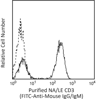
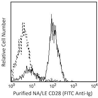
1. Harvest PBMC after stimulation (12 hours) with plate-bound Purified NA/LE Mouse Anti-Human CD3 (Cat. No. 555329) and Purified
NA/LE Mouse Anti-Human CD28 (Cat. No. 555725).
2. Wash the cells twice with stain buffer (eg. BD Pharmingen™ Stain Buffer (FBS), Cat. No. 554656).
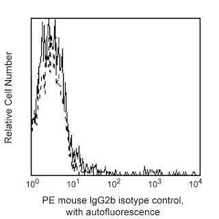
3. Stain 1 × 10^6 cells either with the PE Mouse anti-Human GARP antibody (Cat. No. 562150) or with PE Mouse IgG2b, κ Isotype control
(Cat. No. 556656) for 30 minutes on ice, protected from light.
4. Wash cells twice with stain buffer.
5. Stain with BD Horizon™ V450 Mouse anti-Human FoxP3. Refer to the Technical Data Sheet of Cat. No. 560459 for detailed FoxP3
staining protocol.
In brief,
a. Add 2 ml of 1× FoxP3 buffer A to the cell pellet.
b. Centrifuge and incubate in 0.5 ml of buffer C for 30 minutes.
c. Wash twice with stain buffer and stain with V450 FoxP3 antibody (Cat. No. 560459) for 30-45 minutes.
d. Wash twice with stain buffer and acquire on the Flow cyotmeter.
商品通知
- This reagent has been pre-diluted for use at the recommended Volume per Test. We typically use 1 × 10^6 cells in a 100-µl experimental sample (a test).
- An isotype control should be used at the same concentration as the antibody of interest.
- For fluorochrome spectra and suitable instrument settings, please refer to our Multicolor Flow Cytometry web page at www.bdbiosciences.com/colors.
- Caution: Sodium azide yields highly toxic hydrazoic acid under acidic conditions. Dilute azide compounds in running water before discarding to avoid accumulation of potentially explosive deposits in plumbing.
- Source of all serum proteins is from USDA inspected abattoirs located in the United States.
- BD Horizon V450 has a maximum absorption of 406 nm and maximum emission of 450 nm. Before staining with this reagent, please confirm that your flow cytometer is capable of exciting the fluorochrome and discriminating the resulting fluorescence.
- Please refer to www.bdbiosciences.com/us/s/resources for technical protocols.
配套商品


The 7B11 (also known as CMSSC-7B11) monoclonal antibody specifically binds to human GARP (Glycoprotein A repetitions predominant). The LRRC32 (Leucine rich repeat containing 32) gene encodes the 662 amino acid-residue, 80 kDa transmembrane GARP glycoprotein that has an extracellular region composed primarily of 20 leucine-rich repeats. GARP is specifically expressed on Treg cells activated through the T cell receptor (TCR). Ectopic expression of GARP in human naïve T cells inhibited their proliferation and cytokine secretion upon TCR activation. Remarkably, GARP over-expression in naïve T cells induced expression of FoxP3 and endowed them with a partial suppressive function. The extracellular, but not the cytoplasmic region, of GARP was necessary for these functions. GARP serves as a receptor for latent TGF-beta which may play a role in the suppressive action of Treg cells. GARP is also expressed on platelets and other tissues, however the function on these cells is not known.

研发参考 (2)
-
Ollendorff V, Noguchi T, deLapeyriere O, Birnbaum D. The GARP gene encodes a new member of the family of leucine-rich repeat-containing proteins. Cell Growth Differ. 1994; 5(2):213-219. (Biology). 查看参考
-
Stockis J, Colau D, Coulie PG, Lucas S. Membrane protein GARP is a receptor for latent TGF-beta on the surface of activated human Treg. Eur J Immunol. 2009; 39(12):3315-3322. (Biology). 查看参考
Please refer to Support Documents for Quality Certificates
Global - Refer to manufacturer's instructions for use and related User Manuals and Technical data sheets before using this products as described
Comparisons, where applicable, are made against older BD Technology, manual methods or are general performance claims. Comparisons are not made against non-BD technologies, unless otherwise noted.
For Research Use Only. Not for use in diagnostic or therapeutic procedures.