-
Reagents
- Flow Cytometry Reagents
-
蛋白质印迹试剂
- 免疫分析 试剂
-
Single-Cell Multiomics Reagents
- BD® AbSeq Assay
- BD Rhapsody™ 附件试剂盒
- BD® Single-Cell Multiplexing Kit
- BD Rhapsody™ Targeted mRNA Kits
- BD Rhapsody™ Whole Transcriptome Analysis (WTA) Amplification Kit
- BD Rhapsody™ TCR/BCR Profiling Assays
- BD® OMICS-Guard Sample Preservation Buffer
- BD Rhapsody™ ATAC-Seq Assays
- BD Rhapsody™ TCR/BCR Next Multiomic Assays
-
Functional Assays
-
显微成像试剂
-
Cell Preparation and Separation Reagents
-
- BD® AbSeq Assay
- BD Rhapsody™ 附件试剂盒
- BD® Single-Cell Multiplexing Kit
- BD Rhapsody™ Targeted mRNA Kits
- BD Rhapsody™ Whole Transcriptome Analysis (WTA) Amplification Kit
- BD Rhapsody™ TCR/BCR Profiling Assays
- BD® OMICS-Guard Sample Preservation Buffer
- BD Rhapsody™ ATAC-Seq Assays
- BD Rhapsody™ TCR/BCR Next Multiomic Assays
- China (Chinese)
- 更改国家/语言
Old Browser
Looks like you're visiting us from {countryName}.
Would you like to stay on the current country site or be switched to your country?
Multicolor flow cytometric analysis of CD138 (Syndecan-1) expression on Mouse bone marrow B lymphocytes. C57BL/6 Mouse bone marrow cells were stained with APC Rat Anti-Mouse CD45R/B220 antibody (Cat. No. 553092/561880) and with either BD Horizon™ BV786 Rat IgG2a, κ Isotype Control (Cat. No. 563335; Left Plot) or BD Horizon™ BV786 Rat Anti-Mouse CD138 (Syndecan-1) antibody (Cat. No. 569692; Right Plot) at 0.06 µg/test. BD Via-Probe™ Cell Viability 7-AAD Solution (Cat. No. 555815/555816) was added to cells right before analysis. The bivariate pseudocolor density plot showing the expression of CD138 (Syndecan-1) [or Ig Isotype control staining] versus CD45R/B220 was derived from gated events with the forward and side light-scatter characteristics of viable (7-AAD-negative) bone marrow cells. Flow cytometry and data analysis were performed using a BD LSRFortessa™ X-20 Cell Analyzer System and FlowJo™ Software. Data shown on this Technical Data Sheet are not lot specific.
BD Horizon™ BV786 Rat Anti-Mouse CD138 (Syndecan-1)
监管状态图例
未经BD明确书面授权,严禁使用未经许可的任何商品。
准备和存储
推荐的实验流程
BD® CompBeads can be used as surrogates to assess fluorescence spillover (compensation). When fluorochrome conjugated antibodies are bound to BD® CompBeads, they have spectral properties very similar to cells. However, for some fluorochromes there can be small differences in spectral emissions compared to cells, resulting in spillover values that differ when compared to biological controls. It is strongly recommended that when using a reagent for the first time, users compare the spillover on cells and BD® CompBeads to ensure that BD® CompBeads are appropriate for your specific cellular application.
For optimal and reproducible results, BD Horizon Brilliant Stain Buffer should be used anytime BD Horizon Brilliant dyes are used in a multicolor flow cytometry panel. Fluorescent dye interactions may cause staining artifacts which may affect data interpretation. The BD Horizon Brilliant Stain Buffer was designed to minimize these interactions. When BD Horizon Brilliant Stain Buffer is used in the multicolor panel, it should also be used in the corresponding compensation controls for all dyes to achieve the most accurate compensation. For the most accurate compensation, compensation controls created with either cells or beads should be exposed to BD Horizon Brilliant Stain Buffer for the same length of time as the corresponding multicolor panel. More information can be found in the Technical Data Sheet of the BD Horizon Brilliant Stain Buffer (Cat. No. 563794/566349) or the BD Horizon Brilliant Stain Buffer Plus (Cat. No. 566385).
商品通知
- Please refer to www.bdbiosciences.com/us/s/resources for technical protocols.
- Since applications vary, each investigator should titrate the reagent to obtain optimal results.
- An isotype control should be used at the same concentration as the antibody of interest.
- Caution: Sodium azide yields highly toxic hydrazoic acid under acidic conditions. Dilute azide compounds in running water before discarding to avoid accumulation of potentially explosive deposits in plumbing.
- For fluorochrome spectra and suitable instrument settings, please refer to our Multicolor Flow Cytometry web page at www.bdbiosciences.com/colors.
- Please refer to http://regdocs.bd.com to access safety data sheets (SDS).
- For U.S. patents that may apply, see bd.com/patents.
配套商品
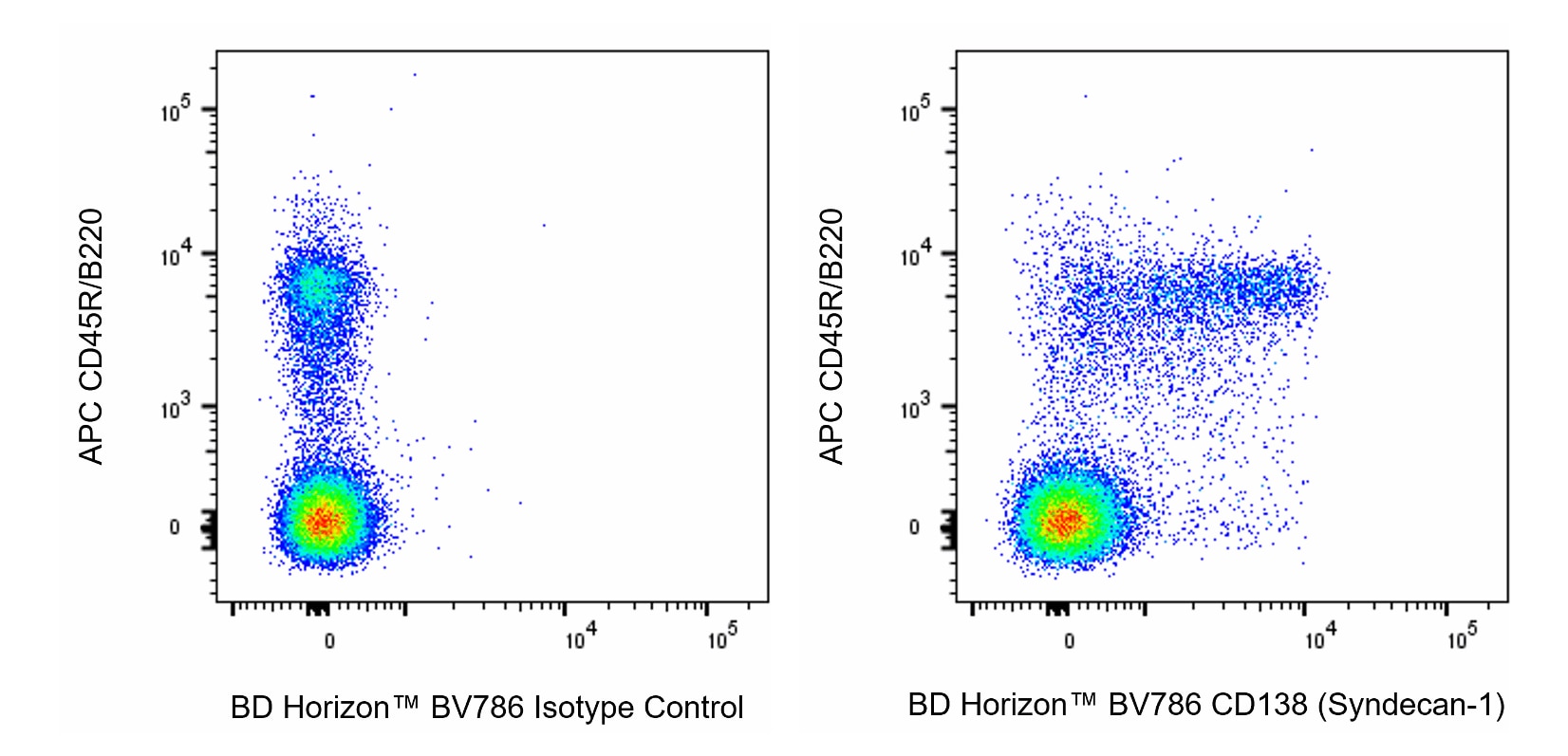
The 281-2 monoclonal antibody specifically binds to the core protein of CD138 (Syndecan-1), a cell-surface, integral membrane heparan sulfate- and chondroitin sulfate-containing proteoglycan that binds to interstitial extracellular matrix molecules. Syndecan-1 is predominantly expressed on epithelial cells, where its expression correlates with normal epithelial organization. It is also expressed on B lymphocytes at specific stages during their differentiation: precursor B cells in the bone marrow, and antibody-secreting cells including plasma cells (but not mature peripheral B cells). It is thus implicated in mediating B cell-matrix interactions. CD138 expression is also regulated during embryonic development, and the molecule shows a tissue- specific structural polymorphism resulting from different post-translational modifications. The 281-2 antibody may be used to detect the differently glycosylated forms, because it reacts with the core protein. Furthermore, the mAb detects the Syndecan-1 ectodomain which is cleaved from cell surfaces by a metalloproteinase.
研发参考 (10)
-
Bernfield M, Kokenyesi R, Kato M, et al. Biology of the syndecans: a family of transmembrane heparan sulfate proteoglycans. Annu Rev Cell Biol. 1992; 8:365-393. (Biology). 查看参考
-
Driver DJ, McHeyzer-Williams LJ, Cool M, Stetson DB, McHeyzer-Williams MG. Development and maintenance of a B220- memory B cell compartment. J Immunol. 2001; 167(3):1393-1405. (Clone-specific: Flow cytometry, Fluorescence microscopy, Immunofluorescence). 查看参考
-
Fitzgerald ML, Wang Z, Park PW, Murphy G, Bernfield M. Shedding of syndecan-1 and -4 ectodomains is regulated by multiple signaling pathways and mediated by a TIMP-3-sensitive metalloproteinase. J Cell Biol. 2000; 148(4):811-824. (Clone-specific: Dot Blot, Fluorescence microscopy, Immunofluorescence). 查看参考
-
Hayashi K, Hayashi M, Jalkanen M, Firestone JH, Trelstad RL, Bernfield M. Immunocytochemistry of cell surface heparan sulfate proteoglycan in mouse tissues. A light and electron microscopic study. J Histochem Cytochem. 1987; 35(10):1079-1088. (Clone-specific: Immunohistochemistry). 查看参考
-
Jalkanen M, Nguyen H, Rapraeger A, Kurn N, Bernfield M. Heparan sulfate proteoglycans from mouse mammary epithelial cells: localization on the cell surface with a monoclonal antibody. J Cell Biol. 1985; 101(3):976-984. (Immunogen: Dot Blot, ELISA, Fluorescence microscopy, Immunofluorescence, Radioimmunoassay, Western blot). 查看参考
-
Lalor PA, Nossal GJ, Sanderson RD, McHeyzer-Williams MG. Functional and molecular characterization of single, (4-hydroxy-3-nitrophenyl)acetyl (NP)-specific, IgG1+ B cells from antibody-secreting and memory B cell pathways in the C57BL/6 immune response to NP. Eur J Immunol. 1992; 22(11):3001-3011. (Biology: Western blot). 查看参考
-
Sanderson RD, Lalor P, Bernfield M. B lymphocytes express and lose syndecan at specific stages of differentiation. Cell Regul. 1989; 1(1):27-35. (Clone-specific: Flow cytometry, Immunoaffinity chromatography, Immunohistochemistry, Western blot). 查看参考
-
Sanderson RD, Sneed TB, Young LA, Sullivan GL, Lander AD. Adhesion of B lymphoid (MPC-11) cells to type I collagen is mediated by integral membrane proteoglycan, syndecan. J Immunol. 1992; 148(12):3902-3911. (Clone-specific: Immunoaffinity chromatography, Radioimmunoassay). 查看参考
-
Saunders S, Jalkanen M, O'Farrell S, Bernfield M. Molecular cloning of syndecan, an integral membrane proteoglycan. J Cell Biol. 1989; 108(4):1547-1556. (Clone-specific). 查看参考
-
Wehrli N, Legler DF, Finke D. Changing responsiveness to chemokines allows medullary plasmablasts to leave lymph nodes. Eur J Immunol. 2001; 31(2):609-616. (Biology: Immunohistochemistry). 查看参考
Please refer to Support Documents for Quality Certificates
Global - Refer to manufacturer's instructions for use and related User Manuals and Technical data sheets before using this products as described
Comparisons, where applicable, are made against older BD Technology, manual methods or are general performance claims. Comparisons are not made against non-BD technologies, unless otherwise noted.
For Research Use Only. Not for use in diagnostic or therapeutic procedures.
Report a Site Issue
This form is intended to help us improve our website experience. For other support, please visit our Contact Us page.