
准备和存储
推荐的实验流程
BD® CompBeads can be used as surrogates to assess fluorescence spillover (compensation). When fluorochrome conjugated antibodies are bound to BD® CompBeads, they have spectral properties very similar to cells. However, for some fluorochromes there can be small differences in spectral emissions compared to cells, resulting in spillover values that differ when compared to biological controls. It is strongly recommended that when using a reagent for the first time, users compare the spillover on cells and BD® CompBeads to ensure that BD® CompBeads are appropriate for your specific cellular application.
For optimal and reproducible results, BD Horizon Brilliant Stain Buffer should be used anytime BD Horizon Brilliant dyes are used in a multicolor flow cytometry panel. Fluorescent dye interactions may cause staining artifacts which may affect data interpretation. The BD Horizon Brilliant Stain Buffer was designed to minimize these interactions. When BD Horizon Brilliant Stain Buffer is used in in the multicolor panel, it should also be used in the corresponding compensation controls for all dyes to achieve the most accurate compensation. For the most accurate compensation, compensation controls created with either cells or beads should be exposed to BD Horizon Brilliant Stain Buffer for the same length of time as the corresponding multicolor panel. More information can be found in the Technical Data Sheet of the BD Horizon Brilliant Stain Buffer (Cat. No. 563794/566349) or the BD Horizon Brilliant Stain Buffer Plus (Cat. No. 566385).
商品通知
- Please refer to www.bdbiosciences.com/us/s/resources for technical protocols.
- This reagent has been pre-diluted for use at the recommended Volume per Test. We typically use 1 × 10^6 cells in a 100-µl experimental sample (a test).
- An isotype control should be used at the same concentration as the antibody of interest.
- Caution: Sodium azide yields highly toxic hydrazoic acid under acidic conditions. Dilute azide compounds in running water before discarding to avoid accumulation of potentially explosive deposits in plumbing.
- For fluorochrome spectra and suitable instrument settings, please refer to our Multicolor Flow Cytometry web page at www.bdbiosciences.com/colors.
- BD Horizon Brilliant Violet 421 is covered by one or more of the following US patents: 8,158,444; 8,362,193; 8,575,303; 8,354,239.
- BD Horizon Brilliant Stain Buffer is covered by one or more of the following US patents: 8,110,673; 8,158,444; 8,575,303; 8,354,239.
- Human donor specific background has been observed in relation to the presence of anti-polyethylene glycol (PEG) antibodies, developed as a result of certain vaccines containing PEG, including some COVID-19 vaccines. We recommend use of BD Horizon Brilliant™ Stain Buffer in your experiments to help mitigate potential background. For more information visit https://www.bdbiosciences.com/en-us/support/product-notices.
- Please refer to http://regdocs.bd.com to access safety data sheets (SDS).
- For U.S. patents that may apply, see bd.com/patents.
配套商品


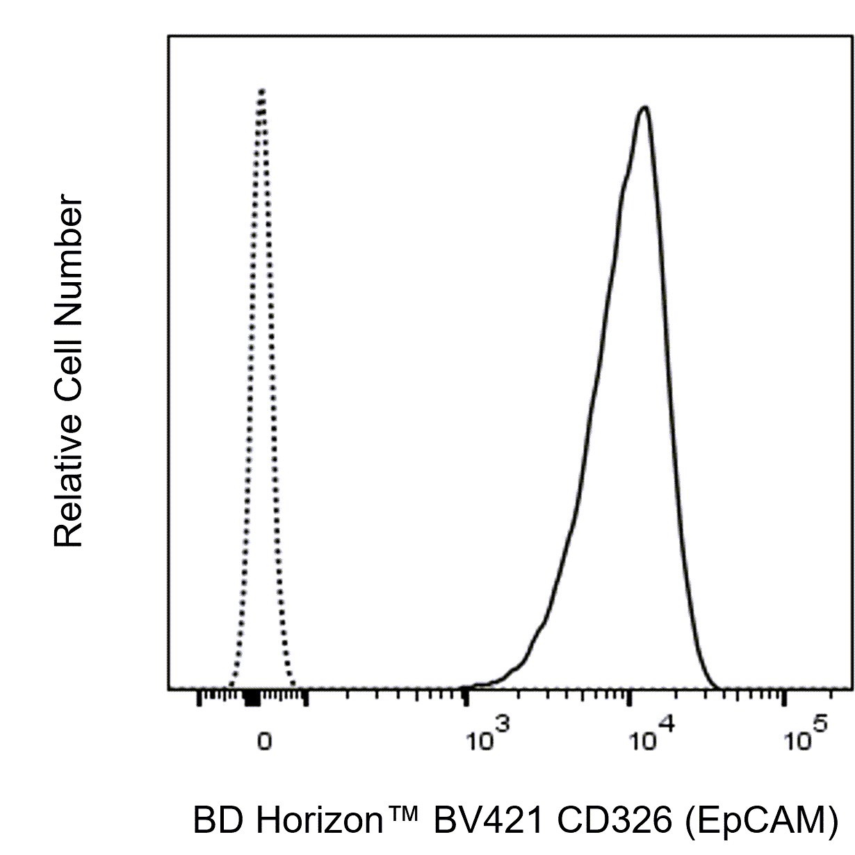
The 9C4 monoclonal antibody specifically binds to human epithelial cell adhesion molecule (EpCAM), also known as adenocarcinoma associated antigen and CD326. EpCAM is an approximately 40-kDa type 1 transmembrane glycoprotein and adhesion molecule that mediates intercellular interactions via homotypic adhesion. The epithelial cells present in non-squamous epithelia and tumors derived from such cells show EpCAM expression. Tumors arising from non-epithelial cells, such as lymphoma, mesothelioma, neuroblastoma, and melanoma, do not express EpCAM. The normal epithelial cells reactive with anti-EpCAM antibodies are those present in the (lower) respiratory tract; the (lower) gastrointestinal tract; tubules in the kidney; the surface epithelium of the ovary; the exocrine and endocrine pancreas; secondary germ cells of telogenic hair follicles; and secretory tubules of sweat glands in the skin, whereas the epidermis is negative. In addition, all epithelial cells in the thyroid and epithelial cells in the thymus show EpCAM expression, while the outer cortex and Hassall's corpuscles have low expression. EpCAM is expressed on a variety of stem and progenitor cells, and its down-regulation is associated with decreased proliferation and differentiation toward endoderm and mesoderm lineages.

研发参考 (5)
-
Bühring HJ, Müller T, Herbst R, et al. The adhesion molecule E-cadherin and a surface antigen recognized by the antibody 9C4 are selectively expressed on erythroid cells of defined maturational stages.. Leukemia. 1996; 10(1):106-16. (Clone-specific: Flow cytometry). 查看参考
-
Lammers R, Giesert C, Grünebach F, Marxer A, Vogel W, Bühring HJ. Monoclonal antibody 9C4 recognizes epithelial cellular adhesion molecule, a cell surface antigen expressed in early steps of erythropoiesis.. Exp Hematol. 2002; 30(6):537-45. (Clone-specific: Flow cytometry). 查看参考
-
Ng VY, Ang SN, Chan JX, Choo AB. Characterization of epithelial cell adhesion molecule as a surface marker on undifferentiated human embryonic stem cells.. Stem Cells. 2010; 28(1):29-35. (Biology). 查看参考
-
Patriarca C, Macchi RM, Marschner AK, Mellstedt H. Epithelial cell adhesion molecule expression (CD326) in cancer: a short review. Cancer Treat Rev. 2012; 38(1):68-75. (Biology). 查看参考
-
Trzpis M, McLaughlin PM, de Leij LM, Harmsen MC. Epithelial cell adhesion molecule: more than a carcinoma marker and adhesion molecule. Am J Pathol. 2007; 171(2):386-395. (Biology). 查看参考
Please refer to Support Documents for Quality Certificates
Global - Refer to manufacturer's instructions for use and related User Manuals and Technical data sheets before using this products as described
Comparisons, where applicable, are made against older BD Technology, manual methods or are general performance claims. Comparisons are not made against non-BD technologies, unless otherwise noted.
For Research Use Only. Not for use in diagnostic or therapeutic procedures.