Skip to main content Skip to navigation
Biotin Mouse Anti-GFP
Figure 1. Flow cytometric analysis of GFP expression in transfected human embryonic kidney cells. AcGFP-transfected HEK-293 cells were fixed with BD Cytofix™ Fixation Buffer (Cat. No. 554655) and permeabilized with the BD Phosflow™ Perm Buffer III (Cat. No. 558050). The cells were then stained with either Biotin Mouse IgG1 κ Isotype Control (dashed line, Cat. No. 555747) or Biotin Mouse Anti-GFP monoclonal antibody (solid line, Cat. No. 565271) followed by APC Streptavidin (Cat. No. 554067). The histograms were derived from gated events based on light scattering characteristics of intact AcGFP-transfected HEK-293 cells. Flow cytometry was performed on a BD LSRFortessa™ Cell Analyzer System. Figure 2. Immunofluorescent staining of GFP-transfected human embryonic kidney cells. Untransfected HEK-293 cells (left panel) and AcGFP-transfected HEK-293 cells (middle and right panels) were fixed with BD Cytofix™ Fixation Buffer (Cat. No. 554655) and permeabilized with 0.1% Triton™ X-100. The cells were blocked with Avidin D followed by Biotin (Vector Labs), and then stained with Biotin Mouse Anti-GFP monoclonal antibody (left and middle panels, pseudo-colored red) at 20 μg/mL. The secondary reagent was Streptavidin Alexa Fluor® 555 (Life Technologies) at 5 μg/mL. GFP signal in the AcGFP-transfected HEK-293 cells is shown for comparison (right panel, pseudo-colored green). Cell nuclei were counterstained with DAPI (Cat. No. 564907). The images were captured on a BD Pathway™ 435 Cell Analyzer and merged using BD Attovision™ software.
商品详情
Down Arrow Up Arrow


BD Pharmingen™
Mouse IgG1, κ
Intracellular staining (flow cytometry) (Routinely Tested), Bioimaging, Immunofluorescence (Tested During Development), Western blot (Not Recommended)
0.5 mg/ml
AB_2739144
Aqueous buffered solution containing ≤0.09% sodium azide.
RUO


准备和存储

Store undiluted at 4°C. The monoclonal antibody was purified from tissue culture supernatant or ascites by affinity chromatography. The antibody was conjugated with biotin under optimum conditions, and unreacted biotin was removed.

商品通知

  1. Since applications vary, each investigator should titrate the reagent to obtain optimal results.
  2. An isotype control should be used at the same concentration as the antibody of interest.
  3. Caution: Sodium azide yields highly toxic hydrazoic acid under acidic conditions. Dilute azide compounds in running water before discarding to avoid accumulation of potentially explosive deposits in plumbing.
  4. Triton is a trademark of the Dow Chemical Company.
  5. For fluorochrome spectra and suitable instrument settings, please refer to our Multicolor Flow Cytometry web page at www.bdbiosciences.com/colors.
  6. Please refer to www.bdbiosciences.com/us/s/resources for technical protocols.
565271 Rev. 1
抗体详情
Down Arrow Up Arrow
1A12-6-18

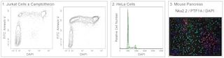
Monoclonal antibody 1A12-6-18 reacts with Green Fluorescent Protein (GFP), which is a 27 kDa bioluminescent protein first purified from the jellyfish, Aequorea victoria. GFP exhibits green fluorescence when exposed to blue or ultraviolet light. Additionally, GFP can be introduced into the genome of and expressed in a wide variety of cell types, making it a useful reporter for tracking gene expression or protein localization. A number of GFP mutants have been developed with varying fluorescence intensity and spectra. Monoclonal antibody 1A12-6-18 is known to react with AcGFP, and is not reactive towards ZsGreen1.

565271 Rev. 1
格式详情
Down Arrow Up Arrow
Biotin
Biotin is a ubiquitous co-factor (also known as Vitamin B7) that has many properties that make it extremely useful for molecular biology. Biotin has an extremely high affinity for the Avidin family of proteins (Kd = 10-15 M), making it the perfect tool to link two molecules. Biotin labeled antibodies can be combined with any number of Avidin-conjugated probes in order to customize an assay to a particular need. This is especially useful in the case of magnetic cell separation using streptavidin/magnetic bead conjugates, or in the case of flow cytometry using streptavidin/fluorophore conjugates.
Biotin
565271 Rev.1
报价单和参考
Down Arrow Up Arrow

研发参考 (4)

  1. Chalfie M, Tu Y, Euskirchen G, Ward WW, Prasher DC. Green fluorescent protein as a marker for gene expression. Science. 1994; 263(5148):802-805. (Biology). 查看参考
  2. Heinen AP, Wanke F, Moos S, et al. Improved Method to Retain Cytosolic Reporter Protein Fluorescence While Staining for Nuclear Proteins. Cytometry A. 2014; 85A:621-627. (Biology). 查看参考
  3. Kain SR, Adams M, Kondepudi A, Yang TT, Ward WW, Kitts P. Green fluorescent protein as a reporter of gene expression and protein localization. Biotechniques. 1995; 19(4):650-655. (Biology). 查看参考
  4. Nybo K. GFP Imaging in Fixed Cells. Biotechniques. 2012; 52(6):359-360. (Biology). 查看参考
查看所有文件 (4) 查看更少内容
565271 Rev. 1

Please refer to Support Documents for Quality Certificates


Global - Refer to manufacturer's instructions for use and related User Manuals and Technical data sheets before using this products as described


Comparisons, where applicable, are made against older BD Technology, manual methods or are general performance claims.  Comparisons are not made against non-BD technologies, unless otherwise noted.

For Research Use Only. Not for use in diagnostic or therapeutic procedures.