准备和存储
推荐的实验流程
BD® CompBeads can be used as surrogates to assess fluorescence spillover (Compensation). When fluorochrome conjugated antibodies are bound to BD® CompBeads, they have spectral properties very similar to cells. However, for some fluorochromes there can be small differences in spectral emissions compared to cells, resulting in spillover values that differ when compared to biological controls. It is strongly recommended that when using a reagent for the first time, users compare the spillover on cells and BD® CompBeads to ensure that BD® CompBeads are appropriate for your specific cellular application.
商品通知
- Since applications vary, each investigator should titrate the reagent to obtain optimal results.
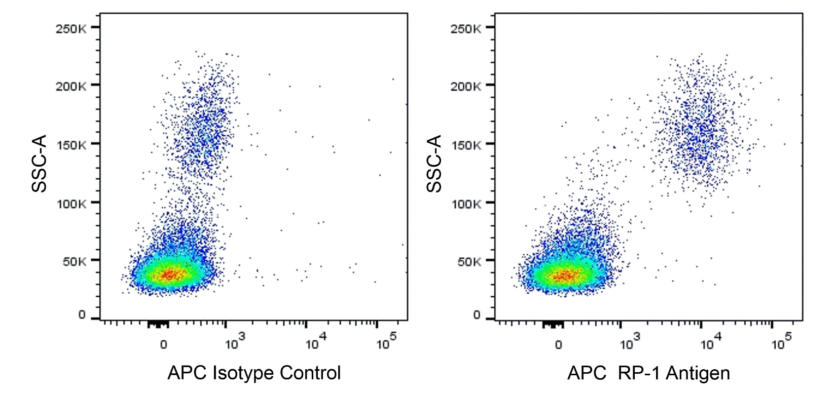
- An isotype control should be used at the same concentration as the antibody of interest.
- Caution: Sodium azide yields highly toxic hydrazoic acid under acidic conditions. Dilute azide compounds in running water before discarding to avoid accumulation of potentially explosive deposits in plumbing.
- This APC-conjugated reagent can be used in any flow cytometer equipped with a dye, HeNe, or red diode laser.
- For fluorochrome spectra and suitable instrument settings, please refer to our Multicolor Flow Cytometry web page at www.bdbiosciences.com/colors.
- Please refer to http://regdocs.bd.com to access safety data sheets (SDS).
- Please refer to www.bdbiosciences.com/us/s/resources for technical protocols.


The RP-1 monoclonal antibody specifically recognizes the RP-1 Antigen. This cell surface marker is expressed on rat peritoneal and peripheral blood neutrophils. Amongst bone marrow cells, the RP-1 Antigen is expressed on band form and mature neutrophils but is not expressed on promyelocytes, myelocytes, and metamyelocytes. The RP-1 antibody does not bind to either rat monocytes, macrophages, eosinophils or to peritoneal neutrophils from mice, rabbits, guinea pigs, or to human peripheral blood neutrophils. Expression of the RP-1 Antigen on rat peritoneal neutrophils is enhanced by cellular stimulation with Phorbol 12-Myristate 13-Acetate (PMA) or Concanavalin A (ConA). Immunoprecipitation and SDS-PAGE analysis of non-treated and PMA-activated rat neutrophil membranes with the RP-1 antibody revealed two main bands of approximately 85 kDa. The RP-1 antibody is also known as the Mouse Anti-Rat Granulocytes antibody.

研发参考 (3)
-
Francis WR, Ireland RE, Spear AM, et al. Flow Cytometric Analysis of Hematopoietic Populations in Rat Bone Marrow. Impact of Trauma and Hemorrhagic Shock.. Cytometry A. 2019; 95(11):1167-1177. (Clone-specific: Flow cytometry, Fluorescence activated cell sorting). 查看参考
-
Gotoh S, Itoh M, Fujii Y, Arai S, Sendo F. Enhancement of the expression of a rat neutrophil-specific cell surface antigen by activation with phorbol myristate acetate and concanavalin A. J Immunol. 1986; 137(2):643-650. (Immunogen: Flow cytometry, Immunoprecipitation, Radioimmunoassay). 查看参考
-
Skrajnar S, Anzur Lasnik M, Bedina Zavec A. A flow cytometric method for determination of the blood neutrophil fraction in rats.. J Am Assoc Lab Anim Sci. 2009; 48(2):152-6. (Clone-specific: Flow cytometry). 查看参考
Please refer to Support Documents for Quality Certificates
Global - Refer to manufacturer's instructions for use and related User Manuals and Technical data sheets before using this products as described
Comparisons, where applicable, are made against older BD Technology, manual methods or are general performance claims. Comparisons are not made against non-BD technologies, unless otherwise noted.
For Research Use Only. Not for use in diagnostic or therapeutic procedures.