
准备和存储
推荐的实验流程
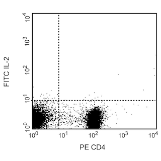
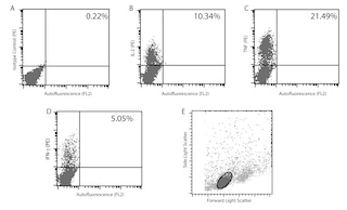
Flow cytometry: The JES6-5H4 antibody is useful for immunofluorescent staining and flow cytometric analysis to identify and enumerate IL-2 producing cells within mixed cell populations. A useful control investigators may consider using for demonstrating specificity of staining, is to pre-block with one of the following reagents: (1) recombinant mouse IL-2 (Cat. No. 550069) or (2) purified JES6-5H4 antibody (Cat. No. 554425), prior to staining.
Cell Preparation: Investigators not wishing to utilize MiCK-1 cells may alternatively prepare mouse splenocytes (e.g BALB/c) stimulated for 4-6 hours with PMA (5 ng/mL, Sigma-Aldrich Cat. No. P-8139) and ionomycin (500 ng/mL, Sigma-Aldrich Cat. No. I-0634) in the presence of
1 µg/mL Brefeldin A (BD GolgiPlug™ Cat. No. 555029). Investigators are advised to fix and permeabilize the cells prior to staining.
商品通知
- Since applications vary, each investigator should titrate the reagent to obtain optimal results.
- An isotype control should be used at the same concentration as the antibody of interest.
- Caution: Sodium azide yields highly toxic hydrazoic acid under acidic conditions. Dilute azide compounds in running water before discarding to avoid accumulation of potentially explosive deposits in plumbing.
- Warning: Some APC-Cy7 and PE-Cy7 conjugates show changes in their emission spectrum with prolonged exposure to formaldehyde. If you are unable to analyze fixed samples within four hours, we recommend that you use BD™ Stabilizing Fixative (Cat. No. 338036).
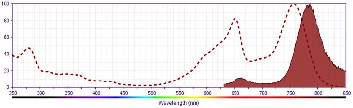
- Please observe the following precautions: Absorption of visible light can significantly alter the energy transfer occurring in any tandem fluorochrome conjugate; therefore, we recommend that special precautions be taken (such as wrapping vials, tubes, or racks in aluminum foil) to prevent exposure of conjugated reagents, including cells stained with those reagents, to room illumination.
- APC-Cy7 tandem fluorochrome emission is collected in a detector for fluorescence wavelengths of 750 nm and higher.
- Cy is a trademark of GE Healthcare.
- APC-Cy7 is a tandem fluorochrome composed of Allophycocyanin (APC), which is excited by laser lines between 595 and 647 nm and serves as an energy donor, coupled to the cyanine dye Cy7™, which acts as an energy acceptor and fluoresces at 780 nm. BD Biosciences Pharmingen has maximized the fluorochrome energy transfer in APC-Cy7, thus maximizing its fluorescence emission intensity, minimizing residual emission from APC, and minimizing required electronic compensation in multilaser-laser flow cytometry systems. Note: Although every effort is made to minimize the lot-to-lot variation in residual emission from APC, it is strongly recommended that every lot be tested for differences in the amount of compensation required and that individual compensation controls are run for each APC-Cy7 conjugate.
- For fluorochrome spectra and suitable instrument settings, please refer to our Multicolor Flow Cytometry web page at www.bdbiosciences.com/colors.
- Please refer to www.bdbiosciences.com/us/s/resources for technical protocols.
配套商品

The JES6-5H4 monoclonal antibody specifically binds to mouse interleukin-2 (IL-2), a multifunctional cytokine that plays pivotal roles in immunity and tolerance. It is produced by activated T cells and affects the activation, growth, proliferation and/or differentiation of various cell types including T and B lymphocytes and their precursors, LAK cells, NK cells, and monocytes/macrophages. IL-2 mediates its biological activities by binding to IL-2 receptor complexes. The intermediate affinity IL-2R is comprised of IL-2Rβ (CD122) and common gamma chain (γc; CD132) subunits, whereas the high-affinity IL-2R is comprised of IL-2Rα (CD25), IL-2Rβ, and γc subunits. The JES6-5H4 monoclonal antibody binds to IL-2 and neutralizes its biological activity.
研发参考 (12)
-
Awatsuji H, Furukawa Y, Nakajima M, Furukawa S, Hayashi K.. Interleukin-2 as a neurotrophic factor for supporting the survival of neurons cultured from various regions of fetal rat brain. J Neurosci Res. 1993; 35(3):305-311. (Biology). 查看参考
-
Fujihashi K, McGhee JR, Beagley KW, et al. Cytokine-specific ELISPOT assay. Single cell analysis of IL-2, IL-4 and IL-6 producing cells. J Immunol Methods. 1993; 160(2):181-189. (Biology). 查看参考
-
Gillis S, Ferm MM, Ou W, Smith KA. T cell growth factor: parameters of production and a quantitative microassay for activity. J Immunol. 1978; 120(6):2027-2032. (Biology). 查看参考
-
Gillis S, Ferm MM, Ou W, Smith KA. T cell growth factor: parameters of production and a quantitative microassay for activity. J Immunol. 1978; 120(6):2027-2032. (Biology). 查看参考
-
Kashima N, Nishi-Takaoka C, Fujita T, et al. Unique structure of murine interleukin-2 as deduced from cloned cDNAs. Nature. 1985; 313(6001):402-404. (Biology). 查看参考
-
Kubo M, Cinader B. Polymorphism of age-related changes in interleukin (IL) production: differential changes of T helper subpopulations, synthesizing IL 2, IL 3 and IL 4. Eur J Immunol. 1990; 20(6):1289-1296. (Biology). 查看参考
-
Mochizuki DY, Watson J, Gillis S. Biochemical separation of interleukin 2. J Immunol Methods. 1980; 39(3):185-201. (Biology). 查看参考
-
Mosmann TR, Cherwinski H, Bond MW, Giedlin MA, Coffman RL. Two types of murine helper T cell clone. I. Definition according to profiles of lymphokine activities and secreted proteins. J Immunol. 1986; 136(7):2348-2357. (Biology). 查看参考
-
Prussin C, Metcalfe DD. Detection of intracytoplasmic cytokine using flow cytometry and directly conjugated anti-cytokine antibodies. J Immunol Methods. 1995; 188(1):117-128. (Methodology: Flow cytometry). 查看参考
-
Sander B, Hoiden I, Andersson U, Moller E, Abrams JS. Similar frequencies and kinetics of cytokine producing cells in murine peripheral blood and spleen. Cytokine detection by immunoassay and intracellular immunostaining. J Immunol Methods. 1993; 166(2):201-214. (Methodology: ELISA). 查看参考
-
Watson J, Mochizuki D. Interleukin 2: a class of T cell growth factors. Immunol Rev. 1980; 51:257-278. (Biology). 查看参考
-
Yokota T, Arai N, Lee F, Rennick D, Mosmann T, Arai K. Use of a cDNA expression vector for isolation of mouse interleukin 2 cDNA clones: expression of T-cell growth-factor activity after transfection of monkey cells. Proc Natl Acad Sci U S A. 1985; 82(1):68-72. (Biology). 查看参考
Please refer to Support Documents for Quality Certificates
Global - Refer to manufacturer's instructions for use and related User Manuals and Technical data sheets before using this products as described
Comparisons, where applicable, are made against older BD Technology, manual methods or are general performance claims. Comparisons are not made against non-BD technologies, unless otherwise noted.
For Research Use Only. Not for use in diagnostic or therapeutic procedures.