-
Reagents
- Flow Cytometry Reagents
-
蛋白质印迹试剂
- 免疫分析 试剂
-
Single-Cell Multiomics Reagents
- BD® AbSeq Assay
- BD Rhapsody™ 附件试剂盒
- BD® Single-Cell Multiplexing Kit
- BD Rhapsody™ TCR/BCR Next Multiomic Assays
- BD Rhapsody™ Targeted mRNA Kits
- BD Rhapsody™ Whole Transcriptome Analysis (WTA) Amplification Kit
- BD OMICS-Guard™ Sample Preservation Buffer
- BD Rhapsody™ ATAC-Seq Assays
- BD® OMICS-One Protein Panels
- BD OMICS-One™ WTA Next Assay
-
Functional Assays
-
显微成像试剂
-
Cell Preparation and Separation Reagents
Old Browser
Looks like you're visiting us from {countryName}.
Would you like to stay on the current location or be switched to your location?
BD Pharmingen™ Alexa Fluor® 647 Rat Anti-Mouse IL-4
克隆 11B11 (RUO)
Expression of IL-4 by stimulated CD4+ and CD4-C57BL/6 spleen cells. Splenocytes from C57BL/6 mice were enriched for CD4+ cells by positive selection using anti-CD4 coated plates (GK1.5,10 µg/ml, Cat. No.553726) for 1 hr at 4°C. Cells were harvested and stimulated with plate-bound anti-mouse CD3 (145-2C11,10 µg/ml, Cat. No. 553057) and soluble anti-CD28 (37.51,2 µg/ml, Cat. No. 553294) antibody in the presence of recombinant mouse IL-2 (10 ng/ml, Cat. No. 550069) and IL-4 (50 ng/ml, Cat. No. 550067) for 2 days. The cells were subsequently washed and expanded in IL-2 and IL-4 for 3 days. Following expansion the cells were washed and stimulated for 4 hrs with PMA (5 ng/ml, Sigma, Cat. No. P-8139) and ionomycin (500 ng, Sigma, P-8139) in the presence of Brefeldin A (GolgiPlug, Cat. No. 555029). Following incubation the cells were harvested and stained with PE-anti-mouse CD4 (Cat. No. 553048) and either rat anti-mouse IL-4 (Alexa Fluor® 647-11B11, Cat. No. 557739) (left panel) or immunoglobulin isotype control (Alexa Fluor® 647-R3-34, Cat. No. 557731) (right panel) by using the BD Pharmingen staining protocol. To demonstrate specificity of staining the binding of Alexa Fluor™ 647 was blocked by the preincubation of the conjugated antibody with molar excess of recombinant mouse IL 4 (0.25 µg, Cat. No. 550067, data not shown) and by preincubation of the fixed/permeabilized cells with an excess of unlabelled 11B11 antibody (5 µg, Cat. No. 554433, data not shown) prior to stainining. The quadarant markers for the bivariate dot plots were set based on the autofluorescence and isotype controls.
监管状态图例
未经BD明确书面授权,严禁使用未经许可的任何商品。
准备和存储
推荐的实验流程
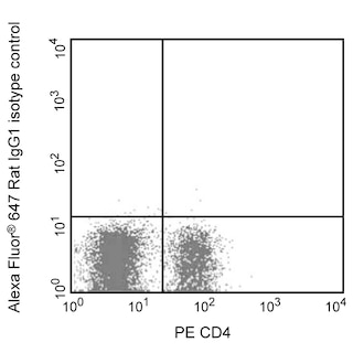
The conjugated 11B11 antibody can be used for multicolor flow cytometric analyses to identify and enumerate IL-4 producing cells within mixed cell populations. For optimal immunofluorescent staining with flow cytometric analysis, this anti-cytokine antibody should be pretitrated. For specific methodology, please visit the protocols section on our web site, www.bdbiosciences.com.
BD® CompBeads can be used as surrogates to assess fluorescence spillover (compensation). When fluorochrome conjugated antibodies are bound to BD® CompBeads, they have spectral properties very similar to cells. However, for some fluorochromes there can be small differences in spectral emissions compared to cells, resulting in spillover values that differ when compared to biological controls. It is strongly recommended that when using a reagent for the first time, users compare the spillover on cell and BD® CompBeads to ensure that BD® CompBeads are appropriate for your specific cellular application.
商品通知
- Since applications vary, each investigator should titrate the reagent to obtain optimal results.
- Please refer to www.bdbiosciences.com/us/s/resources for technical protocols.
- Alexa Fluor® 647 fluorochrome emission is collected at the same instrument settings as for allophycocyanin (APC).
- For fluorochrome spectra and suitable instrument settings, please refer to our Multicolor Flow Cytometry web page at www.bdbiosciences.com/colors.
- The Alexa Fluor®, Pacific Blue™, and Cascade Blue® dye antibody conjugates in this product are sold under license from Molecular Probes, Inc. for research use only, excluding use in combination with microarrays, or as analyte specific reagents. The Alexa Fluor® dyes (except for Alexa Fluor® 430), Pacific Blue™ dye, and Cascade Blue® dye are covered by pending and issued patents.
- Caution: Sodium azide yields highly toxic hydrazoic acid under acidic conditions. Dilute azide compounds in running water before discarding to avoid accumulation of potentially explosive deposits in plumbing.
- This product is provided under an intellectual property license between Life Technologies Corporation and BD Businesses. The purchase of this product conveys to the buyer the non-transferable right to use the purchased amount of the product and components of the product in research conducted by the buyer (whether the buyer is an academic or for-profit entity). The buyer cannot sell or otherwise transfer (a) this product (b) its components or (c) materials made using this product or its components to a third party or otherwise use this product or its components or materials made using this product or its components for Commercial Purposes. Commercial Purposes means any activity by a party for consideration and may include, but is not limited to: (1) use of the product or its components in manufacturing; (2) use of the product or its components to provide a service, information, or data; (3) use of the product or its components for therapeutic, diagnostic or prophylactic purposes; or (4) resale of the product or its components, whether or not such product or its components are resold for use in research. For information on purchasing a license to this product for any other use, contact Life Technologies Corporation, Cell Analysis Business Unit Business Development, 29851 Willow Creek Road, Eugene, OR 97402, USA, Tel: (541) 465-8300. Fax: (541) 335-0504.
- Please refer to http://regdocs.bd.com to access safety data sheets (SDS).
- Alexa Fluor™ is a trademark of Life Technologies Corporation.
- For U.S. patents that may apply, see bd.com/patents.
配套商品
Interleukin-4 (IL-4) is a pleiotropic cytokine that has many roles, such as inducing the differentiation of naïve helper T cells (Th0 cells) to Th2 cells, stimulating activated B-cell and T-cell proliferation, and promoting immunoglobulin class switching to IgG1 and IgE in mouse B-cells. IL-4 is expressed by CD4 T-cells, mast cells, basophils and eosinophils. IL-4 was previously known as B-Cell Differentiation Factor (BCDF) or B-cell Stimulatory Factor (BSF1). The 11B11 monoclonal antibody specifically binds to mouse IL-4. The immunogen used to generate the 11B11 hybridoma was partially purified mouse IL-4 prepared from the supernatant of Phorbol 12-Myristate 13-Acetate (PMA)-stimulated EL-4 cells. The 11B11 antibody is reportedly a neutralizing antibody.
This antibody is routinely tested by flow cytometric analysis. Other applications were tested at BD Biosciences Pharmingen during antibody development only or reported in the literature.
研发参考 (4)
-
Assenmacher M, Schmitz J, Radbruch A. Flow cytometric determination of cytokines in activated murine T helper lymphocytes: expression of interleukin-10 in interferon-gamma and in interleukin-4-expressing cells. Eur J Immunol. 1994; 24(5):1097-1101. (Clone-specific: Flow cytometry). 查看参考
-
Ohara J, Paul WE. Production of a monoclonal antibody to and molecular characterization of B-cell stimulatory factor-1. Nature. 1985; 315(6017):333-336. (Immunogen). 查看参考
-
Sander B, Andersson J, Andersson U. Assessment of cytokines by immunofluorescence and the paraformaldehyde-saponin procedure. Immunol Rev. 1991; 119:65-93. (Clone-specific: ELISA, Flow cytometry). 查看参考
-
Sander B, Hoiden I, Andersson U, Moller E, Abrams JS. Similar frequencies and kinetics of cytokine producing cells in murine peripheral blood and spleen. Cytokine detection by immunoassay and intracellular immunostaining. J Immunol Methods. 1993; 166(2):201-214. (Clone-specific: ELISA, Flow cytometry, Neutralization). 查看参考
Please refer to Support Documents for Quality Certificates
Global - Refer to manufacturer's instructions for use and related User Manuals and Technical data sheets before using this products as described
Comparisons, where applicable, are made against older BD Technology, manual methods or are general performance claims. Comparisons are not made against non-BD technologies, unless otherwise noted.
For Research Use Only. Not for use in diagnostic or therapeutic procedures.