

准备和存储
商品通知
- Since applications vary, each investigator should titrate the reagent to obtain optimal results.
- Please refer to www.bdbiosciences.com/us/s/resources for technical protocols.
- The Alexa Fluor®, Pacific Blue™, and Cascade Blue® dye antibody conjugates in this product are sold under license from Molecular Probes, Inc. for research use only, excluding use in combination with microarrays, or as analyte specific reagents. The Alexa Fluor® dyes (except for Alexa Fluor® 430), Pacific Blue™ dye, and Cascade Blue® dye are covered by pending and issued patents.
- Alexa Fluor® is a registered trademark of Molecular Probes, Inc., Eugene, OR.
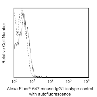
- Alexa Fluor® 647 fluorochrome emission is collected at the same instrument settings as for allophycocyanin (APC).
- Caution: Sodium azide yields highly toxic hydrazoic acid under acidic conditions. Dilute azide compounds in running water before discarding to avoid accumulation of potentially explosive deposits in plumbing.
- For fluorochrome spectra and suitable instrument settings, please refer to our Multicolor Flow Cytometry web page at www.bdbiosciences.com/colors.
- An isotype control should be used at the same concentration as the antibody of interest.
配套商品


The TW7-16B4 monoclonal antibody specifically binds to Latency-Associated Peptide (LAP), a component of the dimeric Transforming Growth Factor-beta 1 (TGF-β1) propeptide encoded by Tgfb1. Prior to secretion, the dimeric LAP-TGF-β1 propeptide is cleaved resulting in a biologically inactive form of dimeric TGF-β1 that is noncovalently associated with dimeric LAP (latent TGF-β1). This complex may be expressed on the surface of TGF-β1-producing cells or be further processed by proteolytic removal of LAP to release the biologically active mature form of the soluble TGF-β1 homodimer. Platelets contain TGF-β1 and most nucleated cells, including tumor cells and cells that comprise the innate and adaptive immune system can produce TGF-β1. TGF-β1 is a potent multifunctional cytokine that regulates numerous processes including development, hematopoiesis, tissue remodeling, wound repair, and immunity as well as cancer and autoimmune diseases.
研发参考 (5)
-
Oida T, Weiner HL. Overexpression of TGF-β1 gene induces cell surface localized glucose-regulated protein 78-associated latency-associated peptide/TGF-β. J Immunol. 2010; 185(6):3529-3535. (Clone-specific). 查看参考
-
Oida T, Weiner HL. TGF-β induces surface LAP expression on murine CD4 T cells independent of Foxp3 induction. PLoS ONE. 2010; 5(11):e15523. (Immunogen: Flow cytometry, Immunoprecipitation, Western blot). 查看参考
-
Oida T, Zhang X, Goto M, et al. CD4+CD25- T cells that express latency-associated peptide on the surface suppress CD4+CD45RBhigh-induced colitis by a TGF-beta-dependent mechanism. J Immunol. 2003; 170(5):2516-2522. (Biology). 查看参考
-
Rubtsov YP, Rudensky AY. TGFbeta signalling in control of T-cell-mediated self-reactivity. Nat Rev Immunol. 2007; 7(6):443-453. (Biology). 查看参考
-
Wilkinson KA, Martin TD, Reba SM, et al. Latency-Associated Peptide of Transforming Growth Factor β enhances mycobacteriocidal immunity in the lung during Mycobacterium bovis BCG infection in C57BL/6 mice. Infect Immun. 2000; 68(11):6505-6508. (Biology). 查看参考
Please refer to Support Documents for Quality Certificates
Global - Refer to manufacturer's instructions for use and related User Manuals and Technical data sheets before using this products as described
Comparisons, where applicable, are made against older BD Technology, manual methods or are general performance claims. Comparisons are not made against non-BD technologies, unless otherwise noted.
For Research Use Only. Not for use in diagnostic or therapeutic procedures.