

准备和存储
推荐的实验流程
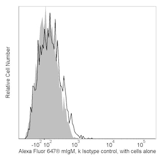
Bioimaging: MN 560850 has been optimized for flow cytometry. For Bioimaging, investigators are encouraged to use MN 560122.
商品通知
- This reagent has been pre-diluted for use at the recommended Volume per Test. We typically use 1 × 10^6 cells in a 100-µl experimental sample (a test).
- An isotype control should be used at the same concentration as the antibody of interest.
- Alexa Fluor® 647 fluorochrome emission is collected at the same instrument settings as for allophycocyanin (APC).
- Alexa Fluor® is a registered trademark of Molecular Probes, Inc., Eugene, OR.
- The Alexa Fluor®, Pacific Blue™, and Cascade Blue® dye antibody conjugates in this product are sold under license from Molecular Probes, Inc. for research use only, excluding use in combination with microarrays, or as analyte specific reagents. The Alexa Fluor® dyes (except for Alexa Fluor® 430), Pacific Blue™ dye, and Cascade Blue® dye are covered by pending and issued patents.
- Caution: Sodium azide yields highly toxic hydrazoic acid under acidic conditions. Dilute azide compounds in running water before discarding to avoid accumulation of potentially explosive deposits in plumbing.
- Source of all serum proteins is from USDA inspected abattoirs located in the United States.
- For fluorochrome spectra and suitable instrument settings, please refer to our Multicolor Flow Cytometry web page at www.bdbiosciences.com/colors.
- Please refer to www.bdbiosciences.com/us/s/resources for technical protocols.
配套商品

The TRA-1-60 monoclonal antibody reacts with the neuraminidase-resistant form of a pluripotent-stem-cell-specific epitope on a high-molecular-weight transmembrane glycoprotein. The TRA-1-60 antigen is a sialylated epitope on the same keratan sulfate core molecule, podocalyxin, as 4 other distinct antigens on tumor-derived cell lines, TRA-1-81, GCTM2, K4, and K21. The expression of TRA-1-60 antigen is stage-specific and can be used to characterize embryonic cells and monitor their differentiation. The antigen is found on teratocarcinoma (embryonal carcinoma or EC), embryonic inner cell mass (but not morula or trophoblast), and embryonic stem (ES) cells. TRA-1-60 antigen is released into the serum of patients bearing testicular tumors containing EC cells. As human EC and ES cells undergo differentiation, expression of TRA-1-60 antigen is lost. Expression of TRA-1-60 antigen has also been observed on a rhesus monkey ES cell line (Thomson et al, 1995).
研发参考 (7)
-
Andrews PW, Banting G, Damanov I, Arnaud D, Avner P. Three monoclonal antibodies defining distinct differentiation antigens associated with different high molecular weight polypeptides on the surface of human embryonal carcinoma cells. Hybridoma. 1984; 3(4):347-361. (Immunogen: Immunofluorescence, Immunoprecipitation, Radioimmunoassay). 查看参考
-
Badcock G, Pigott C, Goepel J, Andrews PW. The human embryonal carcinoma marker antigen TRA-1-60 is a sialylated keratan sulfate proteoglycan. Cancer Res. 1999; 59:4715-4719. (Clone-specific: Immunoprecipitation, Western blot). 查看参考
-
Draper JS, Pigott C, Thomson JA, Andrews PW. Surface antigens of human embryonic stem cells: changes upon differentiation in culture. J Anat. 2002; 200:249-258. (Clone-specific: Flow cytometry). 查看参考
-
Henderson JK, Draper JS, Baillie HS, et al. Preimplantation human embryos and embryonic stem cells show comparable expression of stage-specific embryonic antigens. Stem Cells. 2002; 20:329-337. (Clone-specific: Flow cytometry, Immunofluorescence). 查看参考
-
Schopperle WM, DeWolf WC. The TRA-1-60 and TRA-1-81 human pluripotent stem cell markers are expressed on podocalyxin in embryonal carcinoma. Stem Cells. 2007; 25:723-730. (Clone-specific: Flow cytometry). 查看参考
-
Thomson JA, Itskovitz-Eldor J, Shapiro SS, et al. Embryonic stem cell lines derived from human blastocysts. Science. 1998; 282:1145-1147. (Clone-specific: Immunocytochemistry (cytospins)). 查看参考
-
Thomson JA, Kalishman J, Golos TG, et al. Isolation of a primate embryonic stem cell line. Proc Natl Acad Sci U S A. 1995; 92:7844-7848. (Clone-specific: Immunocytochemistry (cytospins)). 查看参考
Please refer to Support Documents for Quality Certificates
Global - Refer to manufacturer's instructions for use and related User Manuals and Technical data sheets before using this products as described
Comparisons, where applicable, are made against older BD Technology, manual methods or are general performance claims. Comparisons are not made against non-BD technologies, unless otherwise noted.
For Research Use Only. Not for use in diagnostic or therapeutic procedures.