

准备和存储
商品通知
- This reagent has been pre-diluted for use at the recommended Volume per Test. We typically use 1 × 10^6 cells in a 100-µl experimental sample (a test).
- Please refer to www.bdbiosciences.com/us/s/resources for technical protocols.
- The Alexa Fluor®, Pacific Blue™, and Cascade Blue® dye antibody conjugates in this product are sold under license from Molecular Probes, Inc. for research use only, excluding use in combination with microarrays, or as analyte specific reagents. The Alexa Fluor® dyes (except for Alexa Fluor® 430), Pacific Blue™ dye, and Cascade Blue® dye are covered by pending and issued patents.
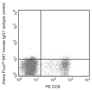
- Alexa Fluor® 647 fluorochrome emission is collected at the same instrument settings as for allophycocyanin (APC).
- Alexa Fluor® is a registered trademark of Molecular Probes, Inc., Eugene, OR.
- Caution: Sodium azide yields highly toxic hydrazoic acid under acidic conditions. Dilute azide compounds in running water before discarding to avoid accumulation of potentially explosive deposits in plumbing.
- For fluorochrome spectra and suitable instrument settings, please refer to our Multicolor Flow Cytometry web page at www.bdbiosciences.com/colors.
- Source of all serum proteins is from USDA inspected abattoirs located in the United States.
- An isotype control should be used at the same concentration as the antibody of interest.
配套商品


The 5.5 monoclonal antibody specifically recognizes S100 calcium binding protein A8 (S100A8) and S100A9 (S100A8/A9), which is also known as Migration inhibitory factor-related protein 8 (MRP-8) and MRP-14 (MRP-8/14), Calprotectin, Cystic fibrosis antigen (CF antigen, CFAG) or L1 light and heavy chain. S100A8 and S100A9 are members of the S100 family that regulate myeloid cell functions, inflammation and innate immune responses. S100A8/A9 (MRP-8/14) is expressed in monocytes and neutrophils. Although not generally expressed in tissue macrophages, S100A8/A9 can be detected within some mononuclear cells comprising inflammatory infiltrates. Soluble S100A8/A9 serves as an early serum marker for detecting and monitoring proinflammatory or immunological disease activities.
研发参考 (6)
-
De Ponti A, Wiechert L, Schneller D, et al. A pro-tumorigenic function of S100A8/A9 in carcinogen-induced hepatocellular carcinoma.. Cancer Lett. 2015; 369(2):396-404. (Biology). 查看参考
-
Hogg N, Allen C, Edgeworth J. Monoclonal antibody 5.5 reacts with p8,14, a myeloid molecule associated with some vascular endothelium.. Eur J Immunol. 1989; 19(6):1053-61. (Immunogen: Immunofluorescence, Immunohistochemistry, Radioimmunoassay). 查看参考
-
Holzinger D, Frosch M, Kastrup A, et al. The Toll-like receptor 4 agonist MRP8/14 protein complex is a sensitive indicator for disease activity and predicts relapses in systemic-onset juvenile idiopathic arthritis.. Ann Rheum Dis. 2012; 71(6):974-80. (Biology). 查看参考
-
Linch DC, Allen C, Beverley PC, Bynoe AG, Scott CS, Hogg N. Monoclonal antibodies differentiating between monocytic and nonmonocytic variants of AML.. Blood. 1984; 63(3):566-73. (Immunogen: Immunocytochemistry, Immunofluorescence). 查看参考
-
Palmer DG, Hogg N, Allen CA, Highton J, Hessian PA. A mononuclear phagocyte subset associated with cell necrosis in rheumatoid nodules: identification with monoclonal antibody 5.5.. Clin Immunol Immunopathol. 1987; 45(1):17-28. (Clone-specific: Immunohistochemistry). 查看参考
-
Sorg C. The calcium binding proteins MRP8 and MRP14 in acute and chronic inflammation.. Behring Inst Mitt. 1992; (91):126-37. (Biology). 查看参考
Please refer to Support Documents for Quality Certificates
Global - Refer to manufacturer's instructions for use and related User Manuals and Technical data sheets before using this products as described
Comparisons, where applicable, are made against older BD Technology, manual methods or are general performance claims. Comparisons are not made against non-BD technologies, unless otherwise noted.
For Research Use Only. Not for use in diagnostic or therapeutic procedures.