-
Reagents
- Flow Cytometry Reagents
-
蛋白质印迹试剂
- 免疫分析 试剂
-
Single-Cell Multiomics Reagents
- BD® AbSeq Assay
- BD Rhapsody™ 附件试剂盒
- BD® Single-Cell Multiplexing Kit
- BD Rhapsody™ Targeted mRNA Kits
- BD Rhapsody™ Whole Transcriptome Analysis (WTA) Amplification Kit
- BD Rhapsody™ TCR/BCR Profiling Assays
- BD® OMICS-Guard Sample Preservation Buffer
- BD Rhapsody™ ATAC-Seq Assays
- BD Rhapsody™ TCR/BCR Next Multiomic Assays
-
Functional Assays
-
显微成像试剂
-
Cell Preparation and Separation Reagents
-
- BD® AbSeq Assay
- BD Rhapsody™ 附件试剂盒
- BD® Single-Cell Multiplexing Kit
- BD Rhapsody™ Targeted mRNA Kits
- BD Rhapsody™ Whole Transcriptome Analysis (WTA) Amplification Kit
- BD Rhapsody™ TCR/BCR Profiling Assays
- BD® OMICS-Guard Sample Preservation Buffer
- BD Rhapsody™ ATAC-Seq Assays
- BD Rhapsody™ TCR/BCR Next Multiomic Assays
- China (Chinese)
-
更改国家/语言
Old Browser
Looks like you're visiting us from {countryName}.
Would you like to stay on the current country site or be switched to your country?


Left Panel - CXCL1 (GROα) expression in human peripheral blood mononuclear cells. PBMCs were cultured without (Left Plots) or with (Right Plots) lipopolysaccharide (LPS) (100 ng/ml, 6 h, 37°C) and BD GolgiStop™ Protein Transport Inhibitor (containing Monensin) (Cat. No. 554724). Cells were harvested, washed, fixed and permeabilized with BD Cytofix/Cytoperm™ Fixation and Permeabilization Solution (Cat. No. 554722). Cells were then washed and stained in BD Perm/Wash™ Buffer (Cat. No. 554723) with BD Horizon™ BV421 Mouse Anti-Human CD14 (Cat. No. 565283) and either Alexa Fluor® 647 Mouse IgG1, κ Isotype Control (Cat. No. 566011; Top Plots) or Alexa Fluor® 647 Mouse Anti-Human CXCL1 (GROα) antibody (Cat. No. 566579; Bottom Plots) at 0.5 ug/test. Two-color flow cytometric contour plots showing the correlated expression of CXCL1 (or Ig Isotype control staining) versus CD14 were derived from gated events with the forward and side light-scatter characteristics of intact cells. Right Panel - CXCL1 (GROα) expression in HiCK-3 Human Cytokine Positive Control Cells. HiCK-3 cells (Cat. No. 555063) were washed and permeabilized using BD Perm/Wash™ Buffer. The cells were stained with BD Horizon BV421 Mouse Anti-Human CD14 and either Alexa Fluor® 647 Mouse IgG1, κ Isotype Control Top Plot) or Alexa Fluor® 647 Mouse Anti-Human CXCL1 (GROα) antibody (Bottom Plot) at 0.5 μg/test. Two-color flow cytometric contour plots showing the correlated expression of CXCL1 (or Ig Isotype control staining) versus CD14 were derived from gated events with the forward and side light-scatter characteristics of intact PBMCs. Flow cytometric analysis was performed using a BD LSRFortessa™ X-20 Cell Analyzer System. Data shown on this Technical Data Sheet are not lot specific.


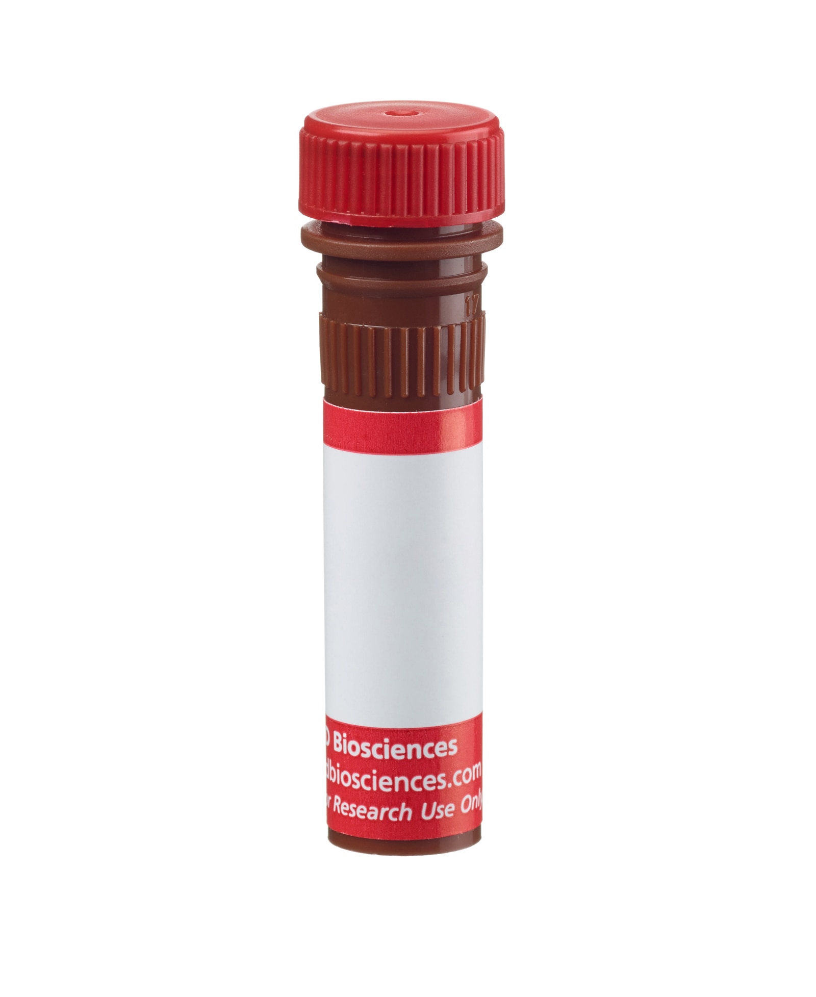
BD Pharmingen™ Alexa Fluor® 647 Mouse Anti-Human CXCL1 (GROα)

监管状态图例
未经BD明确书面授权,严禁使用未经许可的任何商品。
准备和存储
商品通知
- Since applications vary, each investigator should titrate the reagent to obtain optimal results.
- An isotype control should be used at the same concentration as the antibody of interest.
- Caution: Sodium azide yields highly toxic hydrazoic acid under acidic conditions. Dilute azide compounds in running water before discarding to avoid accumulation of potentially explosive deposits in plumbing.
- The Alexa Fluor®, Pacific Blue™, and Cascade Blue® dye antibody conjugates in this product are sold under license from Molecular Probes, Inc. for research use only, excluding use in combination with microarrays, or as analyte specific reagents. The Alexa Fluor® dyes (except for Alexa Fluor® 430), Pacific Blue™ dye, and Cascade Blue® dye are covered by pending and issued patents.
- Alexa Fluor® is a registered trademark of Molecular Probes, Inc., Eugene, OR.
- Alexa Fluor® 647 fluorochrome emission is collected at the same instrument settings as for allophycocyanin (APC).
- For fluorochrome spectra and suitable instrument settings, please refer to our Multicolor Flow Cytometry web page at www.bdbiosciences.com/colors.
- Please refer to www.bdbiosciences.com/us/s/resources for technical protocols.
配套商品


The V40-275 monoclonal antibody specifically recognizes C-X-C motif chemokine ligand 1 (CXCL1) which is also known as Growth-regulated alpha protein (GRO alpha or GROα), Melanoma growth stimulatory activity (MGSA), Fibroblast secretory protein (FSP), Neutrophil-activating protein 3 (NAP-3), or SCYB1. CXCL1 (GROα) is a proinflammatory chemokine that is produced by activated monocytes, macrophages, Kupffer cells, mast cells, fibroblasts, epithelial and endothelial cells in response to proinflammatory stimuli including lipopolysaccharide (LPS), Interleukin-1 (IL-1), or TNF. CXCL1 (GROα) binds to CD182, the CXCR2 chemokine receptor, to attract and activate neutrophils, and to promote angiogenesis and fibrogenesis. Overexpression of CXCL1 (GROα) in some malignant tumors may contribute to tumor vascularization and metastasis.
研发参考 (6)
-
Anisowicz A, Bardwell L, Sager R. Constitutive overexpression of a growth-regulated gene in transformed Chinese hamster and human cells. Proc Natl Acad Sci U S A. 1987; 84(20):7188-7192. (Biology). 查看参考
-
Haskill S, Beg AA, Tompkins SM, et al. Characterization of an immediate-early gene induced in adherent monocytes that encodes I kappa B-like activity. Cell. 1991; 65(7):1281-1289. (Biology). 查看参考
-
Iida N, Grotendorst GR. Cloning and sequencing of a new gro transcript from activated human monocytes: expression in leukocytes and wound tissue. Mol Cell Biol. 1990; 10(10):5596-5599. (Biology). 查看参考
-
Nischalke HD, Berger C, Luda C, et al. The CXCL1 rs4074 A allele is associated with enhanced CXCL1 responses to TLR2 ligands and predisposes to cirrhosis in HCV genotype 1-infected Caucasian patients.. J Hepatol. 2012; 56(4):758-64. (Biology). 查看参考
-
Richmond A, Balentien E, Thomas HG. Molecular characterization and chromosomal mapping of melanoma growth stimulatory activity, a growth factor structurally related to beta-thromboglobulin. EMBO J. 1988; 7(7):2025-2033. (Biology). 查看参考
-
Yang J, Fan GH, Wadzinski BE, Sakurai H, Richmond A. Protein phosphatase 2A interacts with and directly dephosphorylates RelA. J Biol Chem. 2002; 276(51):47828-47833. (Biology). 查看参考
Please refer to Support Documents for Quality Certificates
Global - Refer to manufacturer's instructions for use and related User Manuals and Technical data sheets before using this products as described
Comparisons, where applicable, are made against older BD Technology, manual methods or are general performance claims. Comparisons are not made against non-BD technologies, unless otherwise noted.
For Research Use Only. Not for use in diagnostic or therapeutic procedures.
Report a Site Issue
This form is intended to help us improve our website experience. For other support, please visit our Contact Us page.