Skip to main content Skip to navigation
BUV661 Mouse Anti-Human CD279 (PD-1)
Alert icon
This product is the replacement for 750260.
BUV661 Mouse Anti-Human CD279 (PD-1)
Two-color flow cytometric analysis of CD279 (PD-1) expression on Human peripheral blood lymphocytes. Human whole blood was stained with PE Mouse Anti-Human CD3 antibody (Cat. No. 561809/555333/561808) and with either BD Horizon™ BUV661 Mouse IgG1, κ Isotype Control (Cat. No. 612966; Left Plot) or BD Horizon™ BUV661 Mouse Anti-Human CD279 (PD-1) antibody (Cat. No. 569246; Right Plot) at 1.0 μg/test. The erythrocytes were lysed with BD FACS™ Lysing Solution (Cat. No. 349202). The bivariate pseudocolor density plot showing the correlated expression of CD279 (PD-1) [or Ig Isotype control staining] versus CD3 was derived from gated events with the forward and side light-scatter characteristics of intact lymphocytes. Flow cytometry and data analysis were performed using a BD LSRFortessa™ X-20 Cell Analyzer System and FlowJo™ software. Data shown on this Technical Data Sheet are not lot specific.
商品详情
Down Arrow Up Arrow


BD Horizon™
PD1; hPD-1; hPD-l; PDCD1; PDC1; Programmed cell death 1; SLEB2; hSLE1
Human (QC Testing)
Mouse IgG1, κ
Human PD-1 Recombinant Protein
Flow cytometry (Routinely Tested)
0.2 mg/ml
5133
Aqueous buffered solution containing ≤0.09% sodium azide.
RUO


准备和存储

Store undiluted at 4°C and protected from prolonged exposure to light. Do not freeze. The monoclonal antibody was purified from tissue culture supernatant or ascites by affinity chromatography. The antibody was conjugated to the dye under optimum conditions and unconjugated antibody and free dye were removed.

推荐的实验流程

   BD® CompBeads can be used as surrogates to assess fluorescence spillover (compensation).  When fluorochrome conjugated antibodies are bound to BD® CompBeads, they have spectral properties very similar to cells.   However, for some fluorochromes there can be small differences in spectral emissions compared to cells, resulting in spillover values that differ when compared to biological controls.  It is strongly recommended that when using a reagent for the first time, users compare the spillover on cells and BD® CompBeads to ensure that BD® CompBeads are appropriate for your specific cellular application.

   For optimal and reproducible results, BD Horizon Brilliant Stain Buffer should be used anytime BD Horizon Brilliant dyes are used in a multicolor flow cytometry panel.  Fluorescent dye interactions may cause staining artifacts which may affect data interpretation.  The BD Horizon Brilliant Stain Buffer was designed to minimize these interactions. When BD Horizon Brilliant Stain Buffer is used in in the multicolor panel, it should also be used in the corresponding compensation controls for all dyes to achieve the most accurate compensation. For the most accurate compensation, compensation controls created with either cells or beads should be exposed to BD Horizon Brilliant Stain Buffer for the same length of time as the corresponding multicolor panel. More information can be found in the Technical Data Sheet of the BD Horizon Brilliant Stain Buffer (Cat. No. 563794/566349) or the BD Horizon Brilliant Stain Buffer Plus (Cat. No. 566385).

   Note:  When using high concentrations of antibody, background binding of this dye to erythroid cell subsets (mature erythrocytes and precursors) has been observed.  For researchers studying these cell populations, or in cases where light scatter gating does not adequately exclude these cells from the analysis, this background may be an important factor to consider when selecting reagents for panel(s).

商品通知

  1. Please refer to www.bdbiosciences.com/us/s/resources for technical protocols.
  2. Since applications vary, each investigator should titrate the reagent to obtain optimal results.
  3. An isotype control should be used at the same concentration as the antibody of interest.
  4. Caution: Sodium azide yields highly toxic hydrazoic acid under acidic conditions. Dilute azide compounds in running water before discarding to avoid accumulation of potentially explosive deposits in plumbing.
  5. For fluorochrome spectra and suitable instrument settings, please refer to our Multicolor Flow Cytometry web page at www.bdbiosciences.com/colors.
  6. BD Horizon Brilliant Ultraviolet 661 is covered by one or more of the following US patents: 8,110,673; 8,158,444; 8,227,187; 8,575,303; 8,354,239.
  7. BD Horizon Brilliant Stain Buffer is covered by one or more of the following US patents: 8,110,673; 8,158,444; 8,575,303; 8,354,239.
  8. Human donor specific background has been observed in relation to the presence of anti-polyethylene glycol (PEG) antibodies, developed as a result of certain vaccines containing PEG, including some COVID-19 vaccines. We recommend use of BD Horizon Brilliant™ Stain Buffer in your experiments to help mitigate potential background. For more information visit https://www.bdbiosciences.com/en-us/support/product-notices.
  9. Please refer to http://regdocs.bd.com to access safety data sheets (SDS).
569246 Rev. 1
抗体详情
Down Arrow Up Arrow
EH12.1

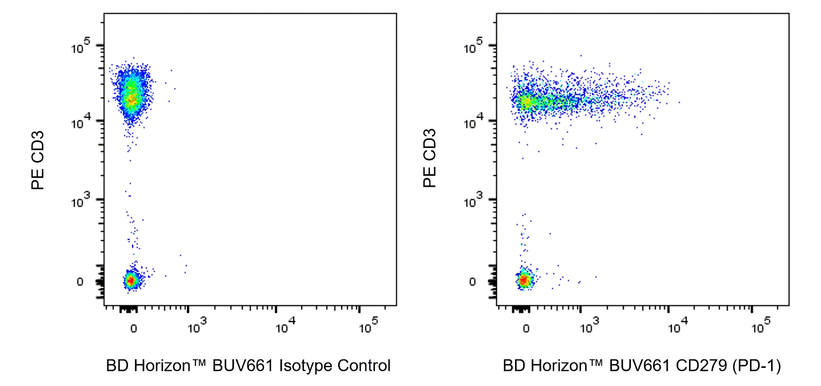
The EH12.1 monoclonal antibody specifically binds to CD279 which is also known as Programmed cell death 1 (PD1). CD279 is an immunoregulatory receptor  expressed on activated T cells, B cells, and myeloid cells. It contains an immunoreceptor tyrosine-based inhibitory motif (ITIM) in the cytoplasmic region. Mice deficient in CD279 show a breakdown of peripheral tolerance and manifest multiple autoimmune symptoms. PD-L1 and PD-L2 are ligands of CD279 and members of the B7 gene family. CD279:PD-Ligands interaction inhibits T cell proliferation and cytokine secretion. Reports suggest that the B7/CTLA-4 pathway primarily attenuates, limits, and/or terminates naïve T-cell activation in secondary lymphoid organs. The PD-ligand:CD279 pathway, on the other hand, may primarily attenuate, limit, and/or terminate T-, B-, and myeloid cell activation/effector function at sites of inflammation in the periphery.

569246 Rev. 1
格式详情
Down Arrow Up Arrow
BUV661
The BD Horizon Brilliant™ Ultraviolet 661 (BUV661) Dye is part of the BD Horizon Brilliant™ Ultraviolet family of dyes. This tandem fluorochrome is comprised of a BUV395 donor with an excitation maximum (Ex Max) of 350-nm and an acceptor dye with an emission maximum (Em Max) at 660-nm. BUV661, driven by BD innovation, is designed to be excited by the ultraviolet laser (355-nm) and detected using an optical filter centered near 660-nm (e.g., 670/25 bandpass filter). The acceptor dye can be excited by the Red (628–640-nm) laser resulting in cross-laser excitation and fluorescence spillover. Please ensure that your instrument’s configurations (lasers and optical filters) are appropriate for this dye.
BUV661
Ultraviolet 355 nm
350 nm
660 nm
569246 Rev.1
报价单和参考
Down Arrow Up Arrow

研发参考 (9)

  1. Bennett F, Luxenberg D, Ling V, et al. Program death-1 engagement upon TCR activation has distinct effects on costimulation and cytokine-driven proliferation: attenuation of ICOS, IL-4, and IL-21, but not CD28, IL-7, and IL-15 responses. J Immunol. 2003; 170(2):711-718. (Biology). 查看参考
  2. Carter L, Fouser LA, Jussif J, et al. PD-1:PD-L inhibitory pathway affects both CD4(+) and CD8(+) T cells and is overcome by IL-2. Eur J Immunol. 2002; 32:634-643. (Biology). 查看参考
  3. Dorfman DM, Brown JA, Shahsafaei A, Freeman GJ. Programmed death-1 (PD-1) is a marker of germinal center-associated T cells and angioimmunoblastic T-cell lymphoma. Am J Surg Pathol. 2006; 30:802-810. (Immunogen: Flow cytometry, Immunohistochemistry). 查看参考
  4. Freeman GJ, Long AJ, Iwai Y, et al. Engagement of PD-1 immunoinhibitory receptor by a novel B7 family member leads to negative regulation of lymphocyte activation. J Exp Med. 2000; 192:1027-1034. (Biology). 查看参考
  5. Kanai T, Totsuka T, Uraushihara K, et al. Blockade of B7-H1 suppresses the development of chronic intestinal inflammation. J Immunol. 2003; 171(8):4156-4163. (Biology). 查看参考
  6. Latchman Y, Wood CR, Chernova T, et al. PD-L2 is a second ligand for PD-1 and inhibits T cell activation. Nat Immunol. 2001; 2(3):261-268. (Biology). 查看参考
  7. Nishimura H, Minato N, Nakano T, Honjo T. Immunological studies on PD-1 deficient mice: implication of PD-1 as a negative regulator for B cell responses. Int Immunol. 1998; 10(10):1563-1572. (Immunogen: Immunoprecipitation). 查看参考
  8. Van der Sluis RM, Kumar NA, Pascoe RD, et al. Combination Immune Checkpoint Blockade to Reverse HIV Latency.. J Immunol. 2020; 204(5):1242-1254. (Clone-specific: Flow cytometry, Fluorescence activated cell sorting). 查看参考
  9. Velu V, Kannanganat S, Ibegbu C, et al. Elevated expression levels of inhibitory receptor programmed death 1 on simian immunodeficiency virus-specific CD8 T cells during chronic infection but not after vaccination. J Virol. 2007; 81(11):5819-5828. (Clone-specific: Blocking, Flow cytometry, Functional assay, Inhibition). 查看参考
查看所有文件 (9) 查看更少内容
569246 Rev. 1

Please refer to Support Documents for Quality Certificates


Global - Refer to manufacturer's instructions for use and related User Manuals and Technical data sheets before using this products as described


Comparisons, where applicable, are made against older BD Technology, manual methods or are general performance claims.  Comparisons are not made against non-BD technologies, unless otherwise noted.

For Research Use Only. Not for use in diagnostic or therapeutic procedures.