The site is undergoing maintenance on
Friday, September 11, 2026, 1:00 - 9:00 am (KST).
If you encounter problems while ordering, please contact us at BDBCustomerService@bd.com
See More
The site is undergoing maintenance on
Friday, September 11, 2026, 1:00 - 9:00 am (KST).
If you encounter problems while ordering, please contact us at BDBCustomerService@bd.com
See MoreThe site is undergoing maintenance on
Friday, September 11, 2026, 1:00 - 9:00 am (KST)
.If you encounter problems while ordering, please contact us at BDBCustomerService@bd.com
Looks like you're visiting us from {countryName}.
Would you like to stay on the current location or be switched to your location?
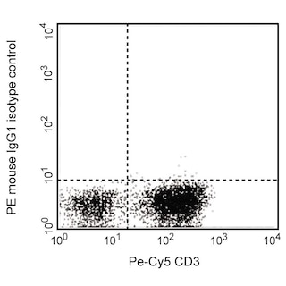
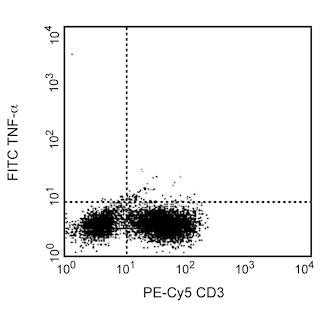
Expression of TNF protein by stimulated human peripheral blood mononuclear cells (PBMC). Human PBMC were stimulated for 6 hours with PMA (50 ng/ml final concentration; Sigma, Cat. #P-8139), calcium ionophore A23187 (0.5 µg/ml final concentration; Sigma, Cat. #C-9275) and PHA (Gibco BRL, Cat. #10576-015) in the presence of GolgiStop™ (2 µM final concentration; Cat. No. 554715). The PBMC were stained with 0.25 µg PE-Cy™5 Mouse Anti-Human CD3 (Cat. No. 555334) and subsequently stained with 0.5 µg of PE Mouse Anti-Human TNF (Cat. No. 554513/557068/559321/562083; left panel). To demonstrate specificity of staining, the binding of PE Mouse Anti-Human TNF was blocked by preincubation of the conjugate with excess (1 µg) recombinant human TNF (Cat. No. 554618; middle panel) and by preincubation of the fixed/permeabilized cells with an excess of Purified Mouse Anti-Human TNF (Cat. No. 554510; right panel) prior to staining with the PE Mouse Anti-Human TNF. The quadrant markers for the bivariate dot plot was set based on the negative staining controls using isotype-matched Ig controls.
Becton, Dickinson and Company의 명시적인 서면 승인 없이는 사용 하실 수 없습니다.
Immunofluorescent Staining and Flow Cytometric Analysis: The MAb11 antibody is useful for intracellular immunofluorescent staining and flow cytometric analysis to identify and enumerate TNF producing cells within mixed cell populations (see image). For optimal immunofluorescent staining with flow cytometric analysis, this anti-cytokine antibody should be pretitrated (≤ 1 µg mAb/million cells). The staining technique and use of blocking controls are described in detail by C. Prussin and D. Metcalfe. For specific methodology, please visit the protocols section under "Cytokines (Intracellular Staining)" or "Intracellular Flow" at our website, http://www.bdbiosciences.com/us/s/resources.
The MAb11 monoclonal antibody specifically binds to human tumor necrosis factor (TNF, also known as TNF-α) protein. TNF is an efficient juxtacrine, paracrine and endocrine mediator of inflammatory and immune functions. It regulates the growth and differentiation of a variety of cell types. TNF is cytotoxic for transformed cells when in conjunction with IFN-γ. It is secreted by activated monocytes/macrophages and other cells such as B cells, T cells and fibroblasts. The immunogen used to generate the MAb11 hybridoma was recombinant human TNF. The MAb11 antibody has been reported to crossreact with Rhesus Macaque TNF.
Please refer to Support Documents for Quality Certificates
Global - Refer to manufacturer's instructions for use and related User Manuals and Technical data sheets before using this products as described
Comparisons, where applicable, are made against older BD Technology, manual methods or are general performance claims. Comparisons are not made against non-BD technologies, unless otherwise noted.
For Research Use Only. Not for use in diagnostic or therapeutic procedures.