


Granzyme B is a serine protease stored primarily in the cytolytic granules of cytotoxic T lymphocytes (CTLs) and natural killer (NK) cells.1 This protein plays a central role in target-cell killing. Our anti-Granzyme B antibody (clone GB11), available in multiple fluorochrome conjugates, enables the identification and characterization of cytotoxic immune cell populations and supports cell-mediated cytotoxicity studies across diverse research applications, including cytotoxicity assays, CAR-T and CAR-NK cell characterization, and tumor-infiltrating lymphocyte (TIL) profiling in human samples.
Applications of Granzyme B staining
T cell cytotoxic potential
Granzyme B is a key mediator and marker of cell-mediated cytotoxicity and is widely used to assess the functional state of cytotoxic lymphocytes. In addition to identifying cytotoxic CD8 T cells, it can help distinguish specialized cytotoxic subsets within the CD4 T-cell compartment, including rare populations of regulatory T cells (Tregs) and follicular helper T cells (Tfh).
For deeper functional profiling, Granzyme B is commonly combined with markers such as perforin, IFN-γ, and CD107a, as well as differentiation markers including CD45RA, CD45RO, CD27, CD62L, and CCR7. Additional markers can be incorporated to characterize specialized populations such as γδ T cells (TCRγδ), Tregs (CD25, CD127, FoxP3), and Tfh cells (CXCR5/CD185). When combined with CAR Detection Reagents, Granzyme B supports research evaluation of the cytotoxic potential of engineered CAR-T cells.

NK (and CAR-NK) cell cytotoxicity
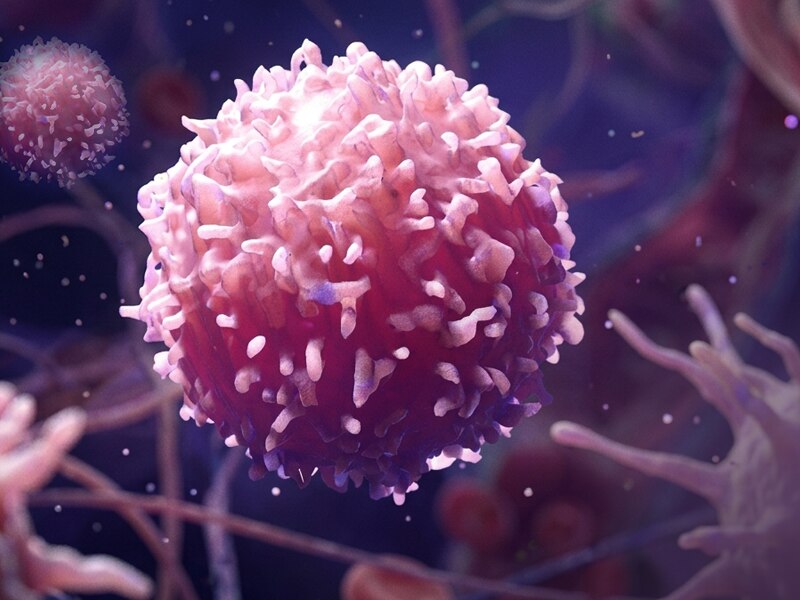
Granzyme B is expressed by mature NK cells, but it is present at higher levels in activated and terminally differentiated populations. Granzyme B expression correlates with cytotoxic capacity and is dynamically regulated by cytokines and target-cell recognition. As a result, Granzyme B expression is often assessed alongside CD56, CD16, and NKG2D. For deeper functional characterization, combine Granzyme B with perforin and (LAMP-1/CD107a) to assess cytotoxic potential, and with CD57 to gain insights into NK-cell maturation and terminal differentiation within research applications.
Tumor microenvironment and immunotherapy response
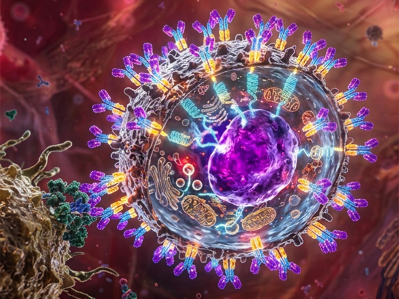
Granzyme B levels are often high in activated tumor-infiltrating lymphocytes (TILs), and low or depleted in exhausted TILs. Therefore, Granzyme B staining is widely used to assess anti-tumor immune responses and monitor immunotherapy efficacy. Panels incorporating Granzyme B often include immune checkpoint markers such as PD-1, TIM-3, LAG-3, and TIGIT, as well as transcription factors such as TOX and TCF-1. Together, these markers provide insight into the activation, functional state, and exhaustion status of lymphocytes in tumor tissues (TILs) and peripheral blood within research applications.
How to stain Granzyme B
Use BD Cytofix/Cytoperm™ kit to fix and permeabilize cells before Granzyme B staining. Because Granzyme B is localized primarily within cytotoxic granules in the cytoplasm, saponin-based permeabilization buffers are particularly well suited for its detection, providing efficient access to intracellular granule-associated proteins while preserving surface marker staining.
Alternatively, BD Phosflow™ Lyse/Fix buffer used in combination with BD Perm/Wash™ Buffer is an effective option when red blood cell lysis is required. Both workflows have been extensively validated for intracellular protein detection by flow cytometry and support robust Granzyme B staining across a variety of immune cell types, including cytotoxic T cells and NK cells.
Considerations for Combined Surface and Granzyme B Staining
When performing surface marker staining in combination with intracellular Granzyme B detection, it is important to consider the impact of fixation and permeabilization on antibody binding and fluorochrome performance. Not all reagents maintain signal integrity following intracellular processing. Therefore, reagent compatibility should be evaluated and, whenever possible, validated under the intended staining conditions before finalizing the panel.
BD Horizon™ Real Dyes are particularly well suited for workflows requiring fixation and permeabilization, as they are designed to provide robust signal stability in different staining conditions. In combination with BD Horizon™ Brilliant Dyes, they can support high-quality multiparametric analysis while preserving sensitivity for both surface and intracellular markers.
In contrast, protein-based fluorochromes such as PE, APC, and especially their tandem conjugates may be more susceptible to changes in signal intensity or spectral profile following fixation and permeabilization.
When using two or more BD Horizon™ Brilliant Dyes, include BD Horizon™ Brilliant Stain Buffer to minimize dye-dye interactions and optimize performance.
Granzyme B Reagents
Browse our Granzyme B antibody portfolio for flow cytometry, featuring clone GB11 in multiple fluorochrome conjugates to support flexible panel design. With demonstrated reactivity to human (QC-tested) and tested in mouse, our portfolio provides the flexibility needed to incorporate Granzyme B staining into a wide range of multicolor assays and supports diverse research applications across immunology, oncology, cell therapy, and drug discovery.
For soluble Granzyme B detection, BD offers both a CBA research assay and an ELISPOT research assay. The BD™ CBA Human Granzyme B Flex Set quantifies secreted Granzyme B in biological fluids and cell culture supernatants, providing a measure of overall cytotoxic activity and enabling multiplex analysis with other soluble immune mediators.
The BD™ Human Granzyme B ELISPOT Set detects individual Granzyme B-secreting cells, making it ideal for evaluating the frequency of functional cytotoxic effector cells in immune monitoring and cell therapy applications.
Product | Research Application | |
560304 (current), previously 558615 | Quantification of soluble human Granzyme B in serum, plasma, and cell culture supernatants. Compatible with multiplex cytokine analysis. | |
558264 (100 tests), 558265 (500 tests) | Required companion reagent kit for running the Human Granzyme B Flex Set. | |
552572 / 563792 (regional numbering varies) | Detects and enumerates individual Granzyme B-secreting cells rather than measuring soluble protein concentration. |
Related Products: support for panel design
As a key indicator of cytotoxic potential, Granzyme B is commonly included in multiparametric immunophenotyping panels to evaluate the functional status of T cells, NK cells, and engineered cell therapies within research applications.
Because Granzyme B is an intracellular protein localized within cytotoxic granules, its detection requires cell fixation and permeabilization. When designing panels, careful planning of reagents for both surface and intracellular staining steps is recommended to ensure workflow compatibility.
Research Application | Markers | Biological Insight |
T Cell and NK Cell Function | Granzyme B, CD3, CD4, CD8, CD56, CD16, IFN-γ, TNF, Perforin, CD107a | Distinguishes T cells from NK cells and assesses cytotoxic function, cytokine production, and degranulation. |
T Cell Exhaustion | Granzyme B, CD3, CD8, PD-1, TIM-3, LAG-3, TIGIT | Measures cytotoxic potential and exhaustion-associated checkpoint expression. |
CAR T Cell Function | Granzyme B, Ki-67, CAR-detection reagent | Assesses cytotoxic potential, proliferation, and confirms CAR-expressing cells. |
NK Cell Function | Granzyme B, CD56, CD16, CD28, CD57 | Correlates NK cell maturation/differentiation states with cytotoxic potential |
T Cell Differentiation | Granzyme B, CD3, CD4, CD8, CD62L, CD27, CD45RA, CD45RO, CCR7 | Correlates Granzyme B expression with naïve, memory, and effector T cell subsets |
Build Your Panel: Visualize in Spectrum Viewer>
Frequently asked questions (FAQs)
1. What is Granzyme B and why is it targeted in flow cytometry?
Granzyme B is a serine protease stored in the cytolytic granules of cytotoxic cells like CTLs and NK cells. It cleaves and activates multiple caspases, gasdermin-E, and other proteins, triggering apoptosis and pyroptosis when inside target cells. As such, it is a marker of cytotoxic immune cell activity and can be used to assess CTL and NK cell effector function, characterize TILs, determine CAR-T cell activity, and check immunotherapy responses within research applications.
2. What fixation and permeabilization protocol is needed for Granzyme B intracellular staining?
As Granzyme B is stored in cytolytic granules within the cytoplasm, use a fixation and permeabilization protocol that enables anti-Granzyme B antibodies to access the cytoplasm. The BD Cytofix/Cytoperm™ kit is validated to fix and permeabilize cells for Granzyme B staining. If red blood cell lysis is needed, the BD Phosflow™ Lyse/Fix buffer together with BD Perm/Wash™ combination is preferred.
3. What is the difference in Granzyme B expression between T cells and NK cells?
Granzyme B is an effector molecule of cytotoxic lymphocytes, and its expression increases upon stimulation. NK cells are typically Granzyme B positive with lower levels on resting cells and higher levels on activated and terminal effector populations. Naive T cells typically show low or no Granzyme B, while activated and effector T cells express it more abundantly, especially CD8+ T cells. Use these distinctions for panel design and positive control selection.
4. What markers should I pair with Granzyme B in a flow cytometry panel?
It depends on the research application. Granzyme B is often paired with CD3/CD4/CD8 for CTL identification, and CD56/CD16 for NK cell studies. For further characterization, it can be combined with markers associated with activation states, as well as specific functional or dysfunctional status. The “Common panels and panel design guidance” section above provides broader panel examples.
5. Which Granzyme B antibody clones and conjugates are available?
Our portfolio includes antibody clones GB11 for flow cytometry and 2C5/F5 for Western Blot. Clone GB11, a Mouse (BALB/c) IgG1 κ isotype with demonstrated reactivity to human and mouse Granzyme B, is conjugated with a broad range of fluorochromes suitable for intracellular staining, including BD Horizon™ Real Dyes, providing optimal performance with fixation and permeabilization protocols.
Related Products
BD Cytofix/Cytoperm™ Fixation/Permeabilization Kit: Use for intracellular Granzyme B staining. This kit contains a fixative and Perm/Wash buffer validated for cytoplasmic protein detection.
BD Pharmingen™ Fixable Viability Stains: Use surface amine-reactive fixable viability markers before fixation in intracellular staining protocols. Our portfolio has multiple options across different laser configurations for flexible panel design.
IFN-γ antibodies: IFN-γ is a commonly used functional co-stain with Granzyme B in CTL and NK activation panels. Both proteins are intracellular, so use the same fixation and permeabilization protocol.
CD8 antibodies: CD8 is the primary lineage marker for CTL identification in panels assessing Granzyme B-mediated cytotoxic function.1 CD8 is a surface marker; stain CD8 before fixation.
References
1. Xie MM, Fang S, Chen Q, Liu H, Wan J, et al. Follicular reglatory T cells inhibit the development of granzyme B-expressing follicular helper T cells. JCI insight; 2019. 10.1172/jci.insight.128076
For Research Use Only. Not for use in diagnostic or therapeutic procedures.