
Extracellular vesicles (EVs) are membranous vesicles that carry tremendous potential as biomarkers and therapeutic targets. Secreted by many cell types, these nanosized particles package diverse biological cargo and play a crucial part in mediating intercellular communication. However, because these intriguing messengers are so small and heterogeneous, they are also difficult to characterize.
Flow cytometry is a common technique for EV analysis1 and is widely used for EV characterization,2,3,4 but there have been several challenges associated with this method. Characterization of EVs using flow cytometry has been challenging owing to their size (30–1,000 nm),1 which is below the detection limit of regular flow cytometers (10–100 µm), and other factors such as the composition of EVs and differences in their concentrations. But innovation and standardization in flow cytometry holds promise for improved methods for their measurement.
BD Biosciences offers instrumentation and resources to tackle some of these challenges. EV researchers have utilized these new advances in innovative ways to resolve several issues that could not be previously addressed.
Read the blog entitled, “Using the latest advances in flow cytometry to tackle challenges in extracellular vesicle characterization”.
Watch the webinar organized by BD Biosciences in collaboration with Nature on the latest developments in extracellular vesicle analysis using flow cytometry with EV researchers Dr. Jonni Moore of Penn Cytomics and Cell Sorting Resource Laboratory, Dr. Terry K. Morgan of Oregon Health & Science University and Dr. Edwin van der Pol of Amsterdam University Medical Centers.
In this webinar, Dr. Edwin van der Pol demonstrates how he used flow cytometry to measure concentrations of EVs that were <100 nm in a reproducible and scalable way, while Dr. Moore and Dr. Morgan discuss mining of EV information from liquid biopsy samples and the detection of gestational phenotype differences using cell-specific EVs, respectively.
This video shows how researchers are using BD products to explore human health and potentially produce future improved treatments and diagnostics.
References
- Gul B, Syed F, Khan S, Iqbal A, Ahmad I. Characterization of extracellular vesicles by flow cytometry: Challenges and promises. Micron. 2022;161:103341. doi: 10.1016/j.micron.2022.103341
- Irmer B, Efing J, Reitnauer LE, et al. Extracellular vesicle-associated tyrosine kinase-like orphan receptors ROR1 and ROR2 promote breast cancer progression. Cell Commun Signal. 2023 ;21(1):171. doi: 10.1186/s12964-023-01186-1
- Mandelbaum N, Zhang L, Carasso S, et al. Extracellular vesicles of the Gram-positive gut symbiont Bifidobacterium longum induce immune-modulatory, anti-inflammatory effects. NPJ Biofilms Microbiomes. 2023;9(1):30. doi: 10.1038/s41522-023-00400-9
- Cohen SJ, Meyerovich G, Blank S, et al. Microbiota transfer following liver surgery involves microbial extracellular vesicle migration that affects liver immunity. Hepatol Commun. 2023;7(6):e0164. doi: 10.1097/HC9.0000000000000164.
A five-step workflow for EV analysis using the BD FACSymphony™ A1 Cell Analyzer, BD FACSDiva™ Software, FlowJo™ Software and Rosetta Calibration Plugin.
Fine tune your small particle detector (SPD)
Step 1: Prepare your instrument using standard small particle beads
- Prepare SPD bead mixture
- Align SPD using the Picomotor Application (if needed)
- Adjust SP SSC voltage to place bead peaks on target

Ensure reproducible low background noise
Step 2: Perform start-up procedure to reduce background noise
- Run BD® Detergent Solution Concentrate
- Run sample diluent and check background noise

Acquire your EV samples and explore the power of “OR” thresholding
Step 3: Apply proper controls, calibration and thresholding strategy
- Use MiFlowCyt-EV framework for publication to guide you on assay controls and SP SSC and fluorescence calibration
- Use “OR” thresholding for SP SSC and relevant fluorescent parameters for maximum information
- Consider running serial dilution series to check for absence of swarming
| 3.1 Buffy only* | 3.5 Single-stained controls* |
| 3.2 Buffer with reagents* | 3.6 Procedural controls* |
| 3.3 Unstained controls* | 3.7 Serial dilution* |
| 3.4 Isotype controls | 3.8 Detergent-treated EV samples |
Welsh JA, et al. Journal of Extracellular Vesicles. 20209;(1):1713526.
Measure Rosetta Calibration beads and your EV samples
STEP 4: Optional for EV size calibration
- Run Rosetta Calibration beads and your EV samples at the same scatter detector voltage and gain
- Configure the threshold and voltage of the scatter channel so that you detect the smallest beads in the mixture while retaining maximum dynamic range
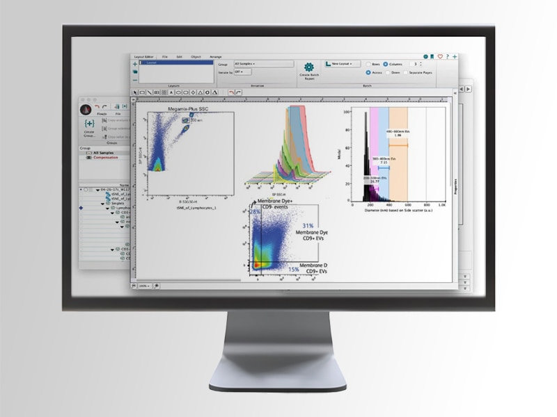
Analyze your EV data
Step 5: Take advantage of the analysis power of BD FACSDiva™ and FlowJo™ Software
Output files from recommended calibration software compatible with BD FACSDiva™ and FlowJo™ Software for further analysis

*Follow routine maintenance process and run CS&T before SPD procedure.
Detection of EVs using the BD FACSymphony™ A1 Cell Analyzer
Resolution of 90-nm polystyrene particles with the BD® Small Particle Detector option

Detection of 90nm polystyrene beads

Separation of 90nm polystyrene beads from noise

Side scatter sensitivity of BD® Small Particle Detector

Characterization of extracellular vesicles from human MCF7 cell line*
* This work was performed in collaboration with Wauben Lab (Utrecht University, The Netherlands) and was supported by the TRAIN-EV Marie Skłodowska-Curie Action-Innovative Training Network, http://train-ev.eu grant agreement No 722148
BD Biosciences offers a robust portfolio of solutions to help you with your extracellular vesicle research.
BD FACSymphony™ A1 Cell Analyzer
The BD FACSymphony™ A1 Cell Analyzer with BD® Small Particle Detector provides a dedicated high-sensitivity detector for a side scatter channel (SP SSC) designed to resolve scatter of small particles such as EVs, viral particle, exosomes and others. This offers the ability to detect particles as small as 90-nm polystyrene beads without changing the instrument setup.

FlowJo™ Software with the Rosetta Calibration plugin
FlowJo™ Software with the Rosetta Calibration plugin, along with the Rosetta Calibration product from Exometry enables calibrated size measurements of small spherical particles (<10 µm).
The plugin allows you to utilize flow cytometry standard (FCS) data captured from Rosetta Calibration beads to derive calibrated size measurements of small particles.* The Rosetta Calibration plugin provides integration between FlowJo™ 10 Software and Rosetta Calibration software.
*The calculation itself is performed by Rosetta Calibration software that needs to be installed separately; please review the Rosetta Calibration manual and download the Rosetta Calibration software from https://www.exometry.com/products/rosetta-calibration (https://www.exometry.com/products/rosetta-calibration).
BD flow cytometers are Class 1 Laser Products.
For Research Use Only. Not for use in diagnostic or therapeutic procedures.