Overview
The BD OMICS-Guard™ Sample Preservation Buffer (SPB) is developed and optimized to address the need for high-quality biological sample preservation over an extended period of time when samples cannot be processed at the same time or location.
- Simple one-step preservation protocol with minimum hands-on time
- Preserves cells for a variety of downstream transcriptomic, epigenetic, proteomic and multiomic applications including RNA-seq, CITE-seq, TCR/BCR profiling, multiomic ATAC-seq,* flow cytometry and qPCR
- Protects cell integrity and preserves different cell populations in your samples for up to 72 hours at 4 °C
- Compatible across multiple sample types: PBMCs, bulk tissue samples and whole blood samples
- Available in two, easy-to-use formats: 50-mL bottle or 12 x 1-mL vials
Interested in long-term sample storage? Check out our BD OMICS-Guard™ CRYO Preservation Buffer
* BD OMICS-Guard™ SPB can be used to preserve single cells prior to nuclei isolation for ATAC-Seq Assays. We do not recommend preserving nuclei samples with BD® OMICS-Guard SPB for ATAC-Seq Assays.

One-Step Sample Preservation Workflow
| Single-cell suspension | Tissue | Whole blood |
Recommended usage | 10,000 to 10 million cells per 1 mL BD OMICS-Guard™ SPB | 30 to 50 mg of tissue per 20 mL BD OMICS-Guard™ SPB | 1:1 Ratio of whole blood (with EDTA) and BD OMICS-Guard™ SPB |
Storage temperature | 4 °C | ||
Storage duration | Up to 72 hours | ||
Buffer handling | BD OMICS-Guard™ SPB should be used with aseptic techniques to prevent contamination of the stock buffer. We recommend working under a laminar flow hood | ||
Sample preservation protocol |
|
|
|
Modifications to single-cell capture on the BD Rhapsody™ Single-Cell Analysis System | For cells/tissues/whole blood preserved in BD OMICS-Guard™ SPB, ensure that the lysis time in the single-cell capture workflow is at least 5 min (can be longer per the specific assay instruction). | ||
Improve Ab-Oligo Signal with the BD® AbSeq Enhancer Kit for BD OMICS-Guard™ SPB-Preserved Samples in CITE-seq
If staining with antibody-oligo cocktails after BD® AbSeq Antibody-Oligos after BD OMICS-Guard™ Buffer preservation, use the BD® AbSeq Enhancer Kit to reduce nonspecific binding events and enhance the AbSeq signal.

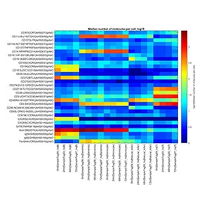
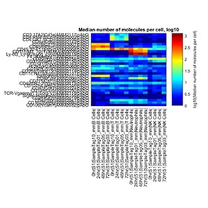
Heatmaps of AbSeq performance with and without the addition of the BD® AbSeq Enhancer Kit. Heatmaps of AbSeq log10(median molecules per cell) expression for different cell types are shown when the BD® AbSeq Enhancer Kit is either not added to the protocol (top) or added to the protocol (bottom) when using preserved cells. Nonspecific background signal (top right) is eliminated, leading to better signal/noise, with the use of the BD® AbSeq Enhancer Kit (bottom right), which closely matches that of the control data (bottom left).
Download the BD OMICS-Guard™ Sample Preservation Buffer product information sheet to access BD® AbSeq Enhancer Kit staining protocols
Applications
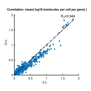
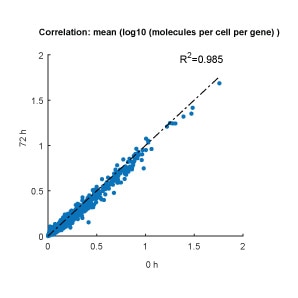
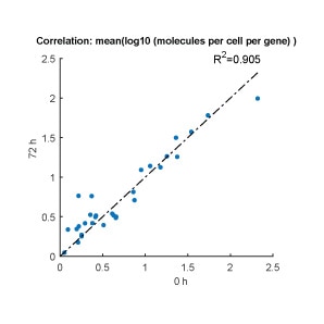
A time course CITE-seq analysis was conducted on the BD Rhapsody™ Single-Cell Analysis System with non-preserved (control) human PBMCs (0 h) and BD OMICS-Guard™ Buffer-preserved human PBMCs at 24, 48 and 72 h. PBMCs at each time point were stained with a 30-plex BD® AbSeq Ab-Oligo Panel. Cell viability, gene expression, surface protein expression and the cell population in the preserved PBMCs at 24, 48 and 72 h were analyzed and compared to that of the control PBMC sample (n = 2)
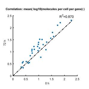
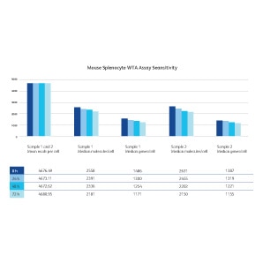
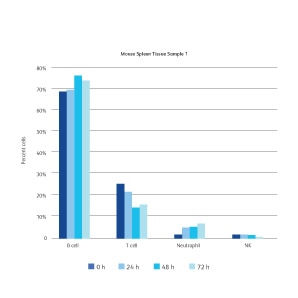
A time course CITE-seq analysis was conducted on the BD Rhapsody™ Single-Cell Analysis System with non-preserved (control) mouse spleen tissues (0 h) and BD OMICS-Guard™ Buffer-preserved mouse spleen tissues at 24, 48 and 72 h. Dissociated mouse splenocytes at each time point were stained with a 30-plex BD® AbSeq Ab-Oligo Panel. Cell viability, gene expression, surface protein expression and the cell population in preserved mouse spleen tissues at 24, 48 and 72 h were analyzed and compared to that of control mouse spleen tissues (n = 2).
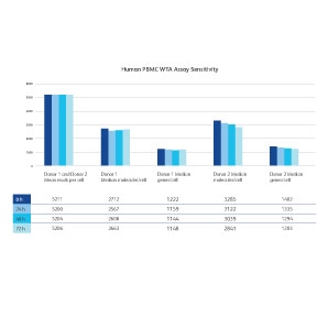
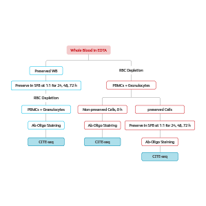
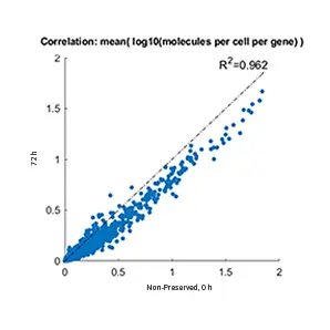
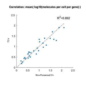
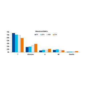
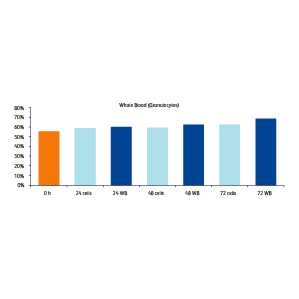
A time course CITE-seq analysis was conducted on the BD Rhapsody™ Single-Cell Analysis System with human whole blood samples collected in EDTA. PBMC and granulocytes were isolated from a portion of the whole blood sample by red blood cell depletion. An aliquot of these cells was immediately processed and not preserved, serving as a 0 h time point. The remaining extracted cells were mixed with BD OMICS-Guard™ SPB and stored at 4 °C for 24, 48, 72 hours (noted in graphs below as “cells”). The rest of the whole blood sample was then directly preserved in BD OMICS-Guard™ SPB by combining the sample with BD OMICS-Guard™ SPB at 1:1 ratio and stored for 24, 48 and 72 h at 4 °C. PBMC and granulocyte isolation was performed by red blood cell depletion at each of the 24, 48 and 72 h preservation time points (noted in graphs below as “WB”). At each time point, cells were stained with the 30-plex BD® OMICS-One Immune Profiler Protein Panel. Whole transcriptome expression, cell surface protein expression and cell type distributions were analyzed at all time points to evaluate the performance of BD OMICS-Guard™ SPB preservation with whole blood.
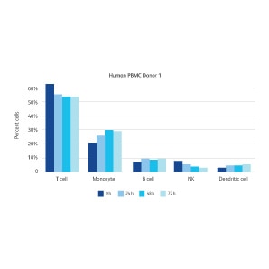
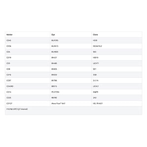
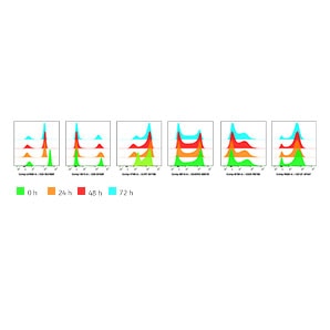
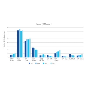
A time course flow cytometry analysis was conducted using non-preserved (control) human PBMCs (0 h) and BD OMICS-Guard™ Buffer-preserved human PBMCs at 24, 48 and 72 h (n = 2). Human PBMCs at each time point were stained with a 13-color fluorescence antibody panel for major immune cell population identification and protein expression analyses and control (0 h) and preserved (24, 48 and 72 h) human PBMCs were compared.
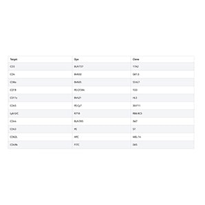
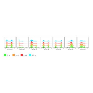
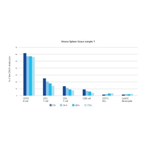
A time-course flow cytometry analysis was conducted using non-preserved (control) mouse spleen tissues (0 h) and BD OMICS-Guard™ Buffer-preserved mouse spleen tissues at 24, 48 and 72 h (n = 2). Dissociated mouse splenocytes at each time point were stained with an 11-color fluorescence antibody panel for major splenic cell population identification and protein expression analyses and control (0 h) and preserved (24, 48 and 72 h) mouse spleen tissues were compared. The protein expression level and frequencies of major cell populations revealed by flow cytometry analyses in mouse splenocytes stayed consistent over the course of 72 hours, indicating the capability of BD OMICS-Guard™ Preservation Buffer to preserve surface protein epitopes and cell composition for flow cytometry analyses.
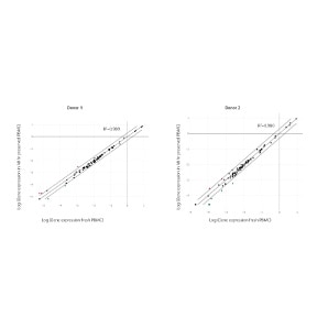
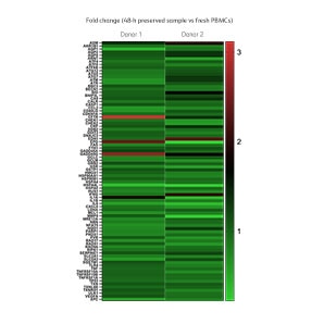
Expression of 84 stress-associated genes were compared between fresh and preserved human PBMC samples using qPCR (n = 2). No significant expression changes of stress-associated genes were observed after 48 h of preservation, indicating a stress-free preservation workflow with BD OMICS-Guard™ Preservation Buffer.
-
Datasheet

-
Product Information Sheet
-
Presentation
-
Protocol
For Research Use Only. Not for use in diagnostic or therapeutic procedures.