

준비 및 보관
제품 고시
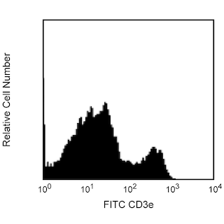
- Since applications vary, each investigator should titrate the reagent to obtain optimal results.
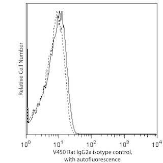
- An isotype control should be used at the same concentration as the antibody of interest.
- Caution: Sodium azide yields highly toxic hydrazoic acid under acidic conditions. Dilute azide compounds in running water before discarding to avoid accumulation of potentially explosive deposits in plumbing.
- For fluorochrome spectra and suitable instrument settings, please refer to our Multicolor Flow Cytometry web page at www.bdbiosciences.com/colors.
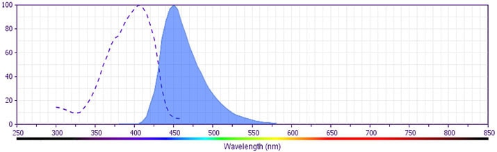
- BD Horizon V450 has a maximum absorption of 406 nm and maximum emission of 450 nm. Before staining with this reagent, please confirm that your flow cytometer is capable of exciting the fluorochrome and discriminating the resulting fluorescence.
- Pacific Blue™ is a trademark of Molecular Probes, Inc., Eugene, OR.
- Please refer to www.bdbiosciences.com/us/s/resources for technical protocols.
관련 제품


The 53-7.3 monoclonal antibody specifically binds to a monomorphic epitope of CD5, a member of the scavenger receptor cysteine-rich protein superfamily and the major ligand of CD72, found on thymocytes, T lymphocytes, thymic NKT cells, and a subset of B lymphocytes, but not on NK cells or splenic NKT cells. The level of surface CD5 expression is developmentally regulated in the thymus, starting with low levels on CD4-CD8- thymocytes and increasing as they mature to CD4+CD8+ then CD4+CD8- or CD4-CD8+ thymocytes. Relatively high levels are maintained on peripheral T lymphocytes. The level of CD5 antigen detected on T helper cells has been reported to be somewhat higher than that on T cytotoxic/suppressor and B cells. Few, if any, intestinal intraepithelial lymphocytes bearing the γδ T-cell receptor express CD5. Phenotypic, anatomical, functional, developmental, and pathogenic characteristics of peripheral CD5+ B cells suggest that they may represent a distinct lineage, known as B-1 cells. The frequency of these CD5+ B cells has been reported to show strain-dependent variation. An additional population of CD5+ B lymphocytes resides in the thymus, where it matures from intrathymic B-cell progenitors. It has been proposed that CD5 is a costimulatory molecule which mediates interactions of cells in the immune system and negatively regulates signal transduction mediated by the T-cell receptor and B-cell receptor.
The antibody is conjugated to BD Horizon™ V450, which has been developed for use in multicolor flow cytometry experiments and is available exclusively from BD Biosciences. It is excited by the Violet laser Ex max of 406 nm and has an Em Max at 450 nm. Conjugates with BD Horizon™ V450 can be used in place of Pacific Blue™ conjugates.
개발 참고 자료 (22)
-
Afar B, Merrill J, Clark EA. Detection of lymphocyte subsets using three-color/single-laser flow cytometry and the fluorescent dye peridinin chlorophyll-alpha protein. J Clin Immunol. 1991; 11(5):254-261. (Methodology: Flow cytometry). 참조 보기
-
Azzam HS, Grinberg A, Lui K, Shen H, Shores EW, Love PE. CD5 expression is developmentally regulated by T cell receptor (TCR) signals and TCR avidity. J Exp Med. 1998; 188(12):2301-2311. (Biology). 참조 보기
-
Bendelac A, Rivera MN, Park SH, Roark JH. Mouse CD1-specific NK1 T cells: development, specificity, and function. Annu Rev Immunol. 1997; 15:535-562. (Biology). 참조 보기
-
Bikah G, Carey J, Ciallella JR, Tarakhovsky A, Bondada S. CD5-mediated negative regulation of antigen receptor-induced growth signals in B-1 B cells. Science. 1996; 274(5294):1906-1909. (Biology). 참조 보기
-
Bikah G, Lynd FM, Aruffo AA, Ledbetter JA, Bondada S. A role for CD5 in cognate interactions between T cells and B cells, and identification of a novel ligand for CD5. Int Immunol. 1998; 10(8):1185-1196. (Biology). 참조 보기
-
Cibotti R, Punt JA, Dash KS, Sharrow SO, Singer A. Surface molecules that drive T cell development in vitro in the absence of thymic epithelium and in the absence of lineage-specific signals. Immunity. 1997; 6(3):245-255. (Biology). 참조 보기
-
Greimers R, Trebak M, Moutschen M, Jacobs N, Boniver J. Improved four-color flow cytometry method using fluo-3 and triple immunofluorescence for analysis of intracellular calcium ion ([Ca2+]i) fluxes among mouse lymph node B- and T-lymphocyte subsets. Cytometry. 1996; 23(3):205-217. (Methodology: Flow cytometry). 참조 보기
-
Hayakawa K, Hardy RR, Parks DR, Herzenberg LA. The "Ly-1 B" cell subpopulation in normal immunodefective, and autoimmune mice. J Exp Med. 1983; 157(1):202-218. (Biology). 참조 보기
-
Hayakawa K, Hardy RR. Normal, autoimmune, and malignant CD5+ B cells: the Ly-1 B lineage. Annu Rev Immunol. 1988; 6:197-218. (Biology). 참조 보기
-
Kantor AB, Herzenberg LA. Origin of murine B cell lineages. Annu Rev Immunol. 1993; 11:501-538. (Biology). 참조 보기
-
Lanier LL. Natural killer cell receptor signaling. Curr Opin Immunol. 2003; 15:308-314. (Biology). 참조 보기
-
Ledbetter JA, Herzenberg LA. Xenogeneic monoclonal antibodies to mouse lymphoid differentiation antigens. Immunol Rev. 1979; 47:63-90. (Immunogen: Immunoprecipitation). 참조 보기
-
Ledbetter JA, Rouse RV, Micklem HS, Herzenberg LA. T cell subsets defined by expression of Lyt-1,2,3 and Thy-1 antigens. Two-parameter immunofluorescence and cytotoxicity analysis with monoclonal antibodies modifies current views. J Exp Med. 1980; 152(2):280-295. (Biology: Cytotoxicity). 참조 보기
-
Lefrancois L. Phenotypic complexity of intraepithelial lymphocytes of the small intestine. J Immunol. 1991; 147(6):1746-1751. (Biology). 참조 보기
-
Luo W, Van de Velde H, von Hoegen I, Parnes JR, Thielemans K. Ly-1 (CD5), a membrane glycoprotein of mouse T lymphocytes and a subset of B cells, is a natural ligand of the B cell surface protein Lyb-2 (CD72). J Immunol. 1992; 148(6):1630-1634. (Biology: ELISA). 참조 보기
-
Masuda K, Makino Y, Cui J, et al. Phenotypes and invariant alpha beta TCR expression of peripheral V alpha 14+ NK T cells. J Immunol. 1997; 158(5):2076-2082. (Biology). 참조 보기
-
Mori S, Inaba M, Sugihara A, et al. Presence of B cell progenitors in the thymus. J Immunol. 1997; 158(9):4193-4199. (Biology). 참조 보기
-
Shapiro HM. Practical Flow Cytometry, 3rd Edition. New York: Wiley-Liss, Inc; 1995:280-281.
-
Tarakhovsky A, Kanner SB, Hombach J, et al. A role for CD5 in TCR-mediated signal transduction and thymocyte selection. Science. 1995; 269(5223):535-537. (Biology). 참조 보기
-
Waggoner AS, Ernst LA, Chen CH, Rechtenwald DJ. PE-CY5. A new fluorescent antibody label for three-color flow cytometry with a single laser. Ann N Y Acad Sci. 1993; 677:185-193. (Methodology: Flow cytometry). 참조 보기
-
Yashiro Y, Tai XG, Toyo-oka K, et al. A fundamental difference in the capacity to induce proliferation of naive T cells between CD28 and other co-stimulatory molecules. Eur J Immunol. 1998; 28(3):926-935. (Biology). 참조 보기
-
van Ewijk W, van Soest PL, van den Engh GJ. Fluorescence analysis and anatomic distribution of mouse T lymphocyte subsets defined by monoclonal antibodies to the antigens Thy-1, Lyt-1, Lyt-2, and T-200. J Immunol. 1981; 127(6):2594-2604. (Biology: Immunohistochemistry). 참조 보기
Please refer to Support Documents for Quality Certificates
Global - Refer to manufacturer's instructions for use and related User Manuals and Technical data sheets before using this products as described
Comparisons, where applicable, are made against older BD Technology, manual methods or are general performance claims. Comparisons are not made against non-BD technologies, unless otherwise noted.
For Research Use Only. Not for use in diagnostic or therapeutic procedures.