-
Reagents
- Flow Cytometry Reagents
-
Western Blotting and Molecular Reagents
- Immunoassay Reagents
-
Single-Cell Multiomics Reagents
- BD® AbSeq Assay
- BD Rhapsody™ Accessory Kits
- BD® Single-Cell Multiplexing Kit
- BD Rhapsody™ Targeted mRNA Kits
- BD Rhapsody™ Whole Transcriptome Analysis (WTA) Amplification Kit
- BD Rhapsody™ TCR/BCR Profiling Assays
- BD OMICS-Guard Sample Preservation Buffer
- BD Rhapsody™ ATAC-Seq Assays
- BD Rhapsody™ TCR/BCR Next Multiomic Assays
-
Functional Assays
-
Microscopy and Imaging Reagents
-
Cell Preparation and Separation Reagents
-
- BD® AbSeq Assay
- BD Rhapsody™ Accessory Kits
- BD® Single-Cell Multiplexing Kit
- BD Rhapsody™ Targeted mRNA Kits
- BD Rhapsody™ Whole Transcriptome Analysis (WTA) Amplification Kit
- BD Rhapsody™ TCR/BCR Profiling Assays
- BD OMICS-Guard Sample Preservation Buffer
- BD Rhapsody™ ATAC-Seq Assays
- BD Rhapsody™ TCR/BCR Next Multiomic Assays
- Korea (Korea)
-
국가 / 언어 변경
Old Browser
Looks like you're visiting us from {countryName}.
Would you like to stay on the current country site or be switched to your country?


Galectin-3 Expression 1. Surface _ PEC: Thioglycolate-elicited C57BL/6 mouse peritoneal exudate cells (PEC) were preincubated with Mouse BD Fc Block™ (Cat. No. 553141/553142) and stained with either Purified Rat IgG2a, κ Isotype Control (Cat. No. 553927; Dotted Line Histogram) or Purified Rat Anti-Galectin-3 (Cat. No. 567905; Solid Line Histogram) at 0.06 µg/test followed by staining with PE Goat Anti-Rat IgG (Cat. No. 550767). DAPI Solution (Cat. No. 564907) was added to cells right before analysis. The histogram shows Galectin-3 expression (or Ig Isotype control staining) derived from gated events with the forward and side light-scatter characteristics of viable (DAPI-negative) cells. 2. Surface + Intracellular _ PEC: Thioglycolate-elicited PEC were fixed with BD Cytofix™ Fixation Buffer (Cat. No. 554655), and permeabilized with BD Perm/Wash™ Buffer (Cat. No. 554723). The cells were stained with either Purified Rat IgG2a, κ Isotype Control (Dotted Line Histogram) or Purified Rat Anti-Galectin-3 (Solid Line Histogram) at 0.06 µg/test, followed by staining with PE Goat Anti-Rat IgG. Total cellular Galectin-3 expression (or Ig Isotype control staining) by intact PEC is shown. Flow cytometry and data analysis were performed using a BD LSRFortessa™ Cell Analyzer and FlowJo™ software. Data shown are not lot specific. 3. Immunohistofluorescence analysis of Galectin-3 expression in mouse intestine. Following antigen retrieval with BD Pharmingen™ Retrievagen A Buffer (Cat. No. 550524), a formalin-fixed, paraffin-embedded BALB/c mouse intestine section was stained with Purified Rat Anti-Galectin-3 (Cat. No. 567904) followed by BD Horizon™ BV480 Goat Anti-Rat Ig (Cat. No. 564878, Left Image; Pseudo-colored Green). Sections were counterstained was with DAPI (Cat. No. 564907; Middle Image; Pseudo-colored Blue) as a nuclear stain. The images were captured and merged (Right Image) using a standard epifluorescence microscope system equipped with a 20× objective.


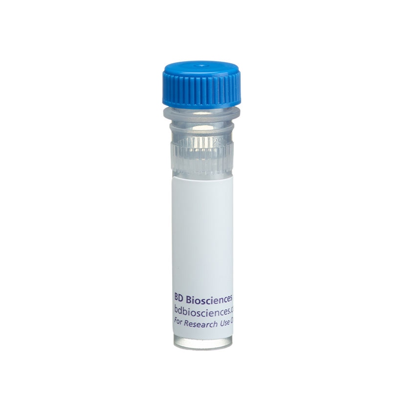
BD Pharmingen™ Purified Rat Anti-Galectin-3

규제 상태 범례
Becton, Dickinson and Company의 명시적인 서면 승인 없이는 사용 하실 수 없습니다.
준비 및 보관
제품 고시
- Please refer to www.bdbiosciences.com/us/s/resources for technical protocols.
- Caution: Sodium azide yields highly toxic hydrazoic acid under acidic conditions. Dilute azide compounds in running water before discarding to avoid accumulation of potentially explosive deposits in plumbing.
- Sodium azide is a reversible inhibitor of oxidative metabolism; therefore, antibody preparations containing this preservative agent must not be used in cell cultures nor injected into animals. Sodium azide may be removed by washing stained cells or plate-bound antibody or dialyzing soluble antibody in sodium azide-free buffer. Since endotoxin may also affect the results of functional studies, we recommend the NA/LE (No Azide/Low Endotoxin) antibody format, if available, for in vitro and in vivo use.
- Please refer to http://regdocs.bd.com to access safety data sheets (SDS).
- Since applications vary, each investigator should titrate the reagent to obtain optimal results.
- An isotype control should be used at the same concentration as the antibody of interest.
관련 제품


.png?imwidth=320)
The M3/38 monoclonal antibody specifically recognizes Galectin-3 (Gal-3 or gal3) which is also known as Galactose-specific lectin 3, Mac-2, MAC2, and Carbohydrate-binding protein 35 (CBP 35). Galectin-3 is an ~30-35 kDa protein that includes an N-terminal proline-rich tandem repeat domain as well as a C-terminal region with one carbohydrate recognition domain (CRDs). Galectin-3 is encoded by Lgals3 (lectin, galactose binding, soluble 3) which belongs to the galectin family. It is expressed by a variety of cells including activated macrophages, dendritic cells (DCs), granulocytes, mast cells, and epithelial cells. Galectin-3 can be detected in monomeric or multimeric forms in cytoplasmic, nuclear, cell surface and extracellular locations. Galectin-3 is a β-galactoside-binding lectin that has multiple intracellular and extracellular functions implicated in biological processes including RNA splicing as well as cellular chemoattraction and adhesion, activation, growth, proliferation, differentiation, and apoptosis. Upregulated Galectin-3 expression is implicated in inflammation and disease states including cancer progression and metastasis.
개발 참고 자료 (8)
-
Demotte N, Wieërs G, Van Der Smissen P, et al. A galectin-3 ligand corrects the impaired function of human CD4 and CD8 tumor-infiltrating lymphocytes and favors tumor rejection in mice.. Cancer Res. 2010; 70(19):7476-88. (Clone-specific: Flow cytometry). 참조 보기
-
Dong R, Zhang M, Hu Q, et al. Galectin-3 as a novel biomarker for disease diagnosis and a target for therapy (Review).. Int J Mol Med. 2018; 41(2):599-614. (Biology). 참조 보기
-
Flotte TJ, Springer TA, Thorbecke GJ. Dendritic cell and macrophage staining by monoclonal antibodies in tissue sections and epidermal sheets. Am J Pathol. 1983; 111(1):112-124. (Clone-specific: Immunohistochemistry). 참조 보기
-
Gray RM, Davis MJ, Ruby KM, Voss PG, Patterson RJ, Wang JL. Distinct effects on splicing of two monoclonal antibodies directed against the amino-terminal domain of galectin-3.. Arch Biochem Biophys. 2008; 475(2):100-8. (Clone-specific: Functional assay, Western blot). 참조 보기
-
Ho MK, Springer TA. Mac-2, a novel 32,000 Mr mouse macrophage subpopulation-specific antigen defined by monoclonal antibodies.. J Immunol. 1982; 128(3):1221-8. (Immunogen: Flow cytometry, Immunoprecipitation, Radioimmunoassay). 참조 보기
-
Johannes L, Jacob R, Leffler H. Galectins at a glance.. J Cell Sci. 2018; 131(9):jcs208884. (Biology). 참조 보기
-
Karlsson A, Follin P, Leffler H, Dahlgren C. Galectin-3 activates the NADPH-oxidase in exudated but not peripheral blood neutrophils.. Blood. 1998; 91(9):3430-8. (Clone-specific: Flow cytometry). 참조 보기
-
Newlaczyl AU, Yu LG. Galectin-3--a jack-of-all-trades in cancer.. Cancer Lett. 2011; 313(2):123-8. (Biology). 참조 보기
Please refer to Support Documents for Quality Certificates
Global - Refer to manufacturer's instructions for use and related User Manuals and Technical data sheets before using this products as described
Comparisons, where applicable, are made against older BD Technology, manual methods or are general performance claims. Comparisons are not made against non-BD technologies, unless otherwise noted.
For Research Use Only. Not for use in diagnostic or therapeutic procedures.
Report a Site Issue
This form is intended to help us improve our website experience. For other support, please visit our Contact Us page.