

준비 및 보관
권장 분석 절차
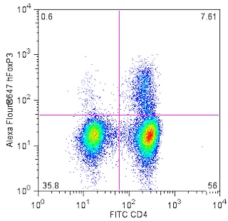
Suggested Staining Procedures for PerCP-Cy™5.5 Mouse Anti-Human LAP antibody:
1. Harvest PBMCs after stimulation (24 hours) with plate-bound Purified NA/LE Mouse Anti-Human CD3 (Cat. No. 555329) and Purified NA/LE Mouse Anti-Human CD28 (Cat. No. 555725) antibodies.
2. Wash the cells twice with stain buffer [eg. BD Pharmingen™ Stain Buffer (FBS), Cat. No. 554656].
3. Stain 1 × 10^6 cells with FITC Mouse Anti-Human CD4 (Cat. No. 340133) and either PerCP-Cy™5.5 Mouse IgG1, κ Isotype Control (Cat. No. 550795) or PerCP-Cy™5.5 Mouse Anti-Human LAP (Cat. No. 562544) antibodies for 30 minutes on ice, protected from light.
4. Wash cells twice with stain buffer.
5. Stain cells for intracellular FoxP3 with Alexa Fluor® 647 Mouse Anti-Human FoxP3 antibody; refer to the Technical Data Sheets of Cat. No. 560889 or 560045 for a detailed protocol.
In brief,
a. Add 2 ml of 1 × FoxP3 buffer A to the cell pellet.
b. Centrifuge and incubate in 0.5 ml of buffer C for 30 minutes.
c. Wash cells twice with stain buffer and stain with fluorescent Anti-FoxP3 antibody for 30-45min.
d. Wash cells twice with stain buffer and analyze by flow cytometry.
제품 고시
- This reagent has been pre-diluted for use at the recommended Volume per Test. We typically use 1 × 10^6 cells in a 100-µl experimental sample (a test).
- Source of all serum proteins is from USDA inspected abattoirs located in the United States.
- An isotype control should be used at the same concentration as the antibody of interest.
- Please refer to www.bdbiosciences.com/us/s/resources for technical protocols.
- Cy is a trademark of Amersham Biosciences Limited. This conjugated product is sold under license to the following patents: US Patent Nos. 5,486,616; 5,569,587; 5,569,766; 5,627,027.
- Please observe the following precautions: Absorption of visible light can significantly alter the energy transfer occurring in any tandem fluorochrome conjugate; therefore, we recommend that special precautions be taken (such as wrapping vials, tubes, or racks in aluminum foil) to prevent exposure of conjugated reagents, including cells stained with those reagents, to room illumination.
- Caution: Sodium azide yields highly toxic hydrazoic acid under acidic conditions. Dilute azide compounds in running water before discarding to avoid accumulation of potentially explosive deposits in plumbing.
- PerCP-Cy5.5–labelled antibodies can be used with FITC- and R-PE–labelled reagents in single-laser flow cytometers with no significant spectral overlap of PerCP-Cy5.5, FITC, and R-PE fluorescence.
- PerCP-Cy5.5 is optimized for use with a single argon ion laser emitting 488-nm light. Because of the broad absorption spectrum of the tandem fluorochrome, extra care must be taken when using dual-laser cytometers, which may directly excite both PerCP and Cy5.5™. We recommend the use of cross-beam compensation during data acquisition or software compensation during data analysis.
- For fluorochrome spectra and suitable instrument settings, please refer to our Multicolor Flow Cytometry web page at www.bdbiosciences.com/colors.
- This product is subject to proprietary rights of Amersham Biosciences Corp. and Carnegie Mellon University and made and sold under license from Amersham Biosciences Corp. This product is licensed for sale only for research. It is not licensed for any other use. If you require a commercial license to use this product and do not have one return this material, unopened to BD Biosciences, 10975 Torreyana Rd, San Diego, CA 92121 and any money paid for the material will be refunded.
관련 제품

The TW4-2F8 monoclonal antibody specifically binds to Latency-Associated Peptide (LAP), a component of the dimeric Transforming Growth Factor-beta 1 (TGF-β1) propeptide encoded by TGFB1. Prior to secretion, the dimeric LAP-TGF-β1 propeptide is cleaved resulting in a biologically inactive form of dimeric TGF-β1 that is noncovalently associated with dimeric LAP (latent TGF-β1). This complex may be expressed on the surface of TGF-β1-producing cells or be further processed by proteolytic removal of LAP to release the biologically active mature form of the soluble TGF-β1 homodimer. Platelets contain TGF-β1 and most nucleated cells, including tumor cells and cells that comprise the innate and adaptive immune system can produce TGF-β1. TGF-β1 is a potent multifunctional cytokine that regulates numerous processes including development, hematopoiesis, tissue remodeling, wound repair, and immunity as well as cancer and autoimmune diseases. Clone TW4-2F8 is routinely quality tested through intracellular staining of LAP in P3UI-TGFβ1 transfected cells.

개발 참고 자료 (2)
-
Oida T, Weiner HL. Overexpression of TGF-β1 gene induces cell surface localized glucose-regulated protein 78-associated latency-associated peptide/TGF-β. J Immunol. 2010; 185(6):3529-3535. (Clone-specific: Flow cytometry, Immunoprecipitation, Western blot). 참조 보기
-
Rubtsov YP, Rudensky AY. TGFbeta signalling in control of T-cell-mediated self-reactivity. Nat Rev Immunol. 2007; 7(6):443-453. (Biology). 참조 보기
Please refer to Support Documents for Quality Certificates
Global - Refer to manufacturer's instructions for use and related User Manuals and Technical data sheets before using this products as described
Comparisons, where applicable, are made against older BD Technology, manual methods or are general performance claims. Comparisons are not made against non-BD technologies, unless otherwise noted.
For Research Use Only. Not for use in diagnostic or therapeutic procedures.