준비 및 보관
제품 고시
- Since applications vary, each investigator should titrate the reagent to obtain optimal results.
- An isotype control should be used at the same concentration as the antibody of interest.
- Caution: Sodium azide yields highly toxic hydrazoic acid under acidic conditions. Dilute azide compounds in running water before discarding to avoid accumulation of potentially explosive deposits in plumbing.
- For fluorochrome spectra and suitable instrument settings, please refer to our Multicolor Flow Cytometry web page at www.bdbiosciences.com/colors.
- Please refer to www.bdbiosciences.com/us/s/resources for technical protocols.
관련 제품


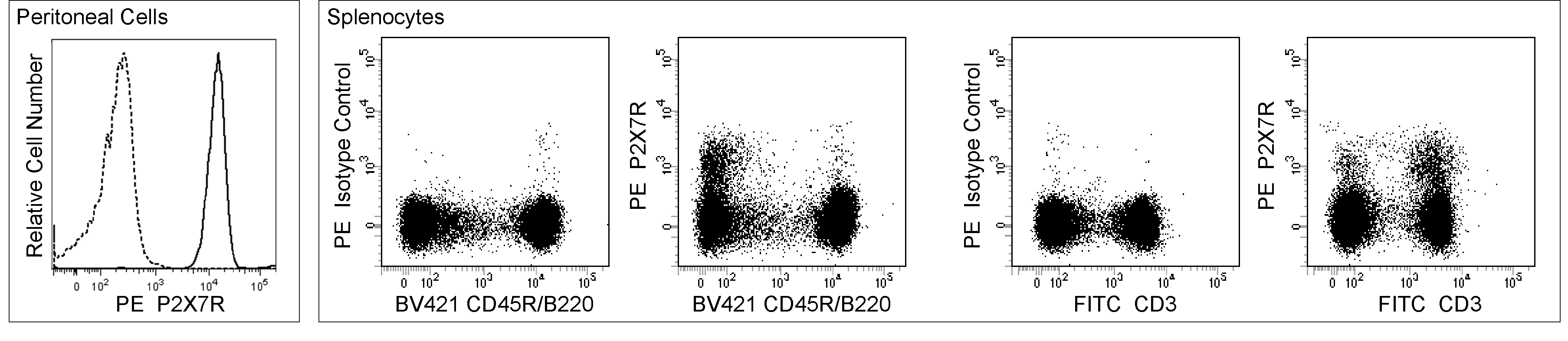
The 1F11monoclonal antibody specifically binds to the P2X7 receptor. P2X7 is also known as the P2X7 Receptor (P2X7R), Purinergic receptor P2X ligand-gated ion channel 7 (P2RX7), P2X7 purinoceptor, P2Z receptor, or ATP receptor. P2X7 is differentially expressed on a wide variety of cell types including T cells, B cells, macrophages, dendritic cells, mast cells, neurons, microglia, and astrocytes. Functional P2X receptors exist as trimers. Each P2X7 monomer contains intracellular N and C termini, two transmembrane domains, and a large extra cellular loop. When present at relatively high levels, extracellular adenosine 5'-triphosphate (ATP) binds to the P2X7 receptor and opens a channel for extracellular cation (eg, Na+, K+, Ca++) influx into cells. The P2X7 receptor-mediated cation influx can cause membrane depolarization and Ca++ mediated signaling cascades that trigger cellular responses, eg, the production and/or release of proinflammatory mediators (eg, IL-1β or IL-18) by cells of the immune system. P2X7 is also implicated in mediating ATP-induced apoptosis of cells. The 1F11 antibody reportedly blocks ATP-mediated cellular activation through P2X7R.

개발 참고 자료 (4)
-
Bulanova E, Bulfone-Paus S. P2 receptor-mediated signaling in mast cell biology. Purinergic Signal. 2010; 6(1):3-17. (Biology). 참조 보기
-
Cloning and functional characterisation of the mouse P2X7 receptor. Cloning and functional characterisation of the mouse P2X7 receptor. FEBS Lett. 1998; 439(1-2):26-30. (Biology). 참조 보기
-
Idzko M, Ferrari D, Eltzschig HK. Nucleotide signalling during inflammation. Nature. 2014; 509(7500):310-317. (Biology).
-
Kurashima Y, Amiya T, Nochi T, et al. Extracellular ATP mediates mast cell-dependent intestinal inflammation through P2X7 purinoceptors.. Nat Commun. 2012; 3:1034. (Immunogen: Functional assay, Immunoprecipitation, Inhibition, In vivo exacerbation). 참조 보기
Please refer to Support Documents for Quality Certificates
Global - Refer to manufacturer's instructions for use and related User Manuals and Technical data sheets before using this products as described
Comparisons, where applicable, are made against older BD Technology, manual methods or are general performance claims. Comparisons are not made against non-BD technologies, unless otherwise noted.
For Research Use Only. Not for use in diagnostic or therapeutic procedures.