준비 및 보관
권장 분석 절차
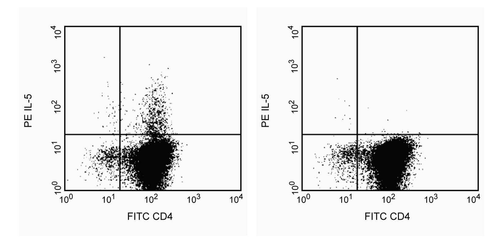
For optimal immunofluorescent staining with flow cytometric analysis, this anti-cytokine antibody should be titrated (≤ 0.5 µg mAb/million cells). A suitable rat IgG1 isotype control assessing the level of background staining on fixed/permeabilized or human cells is PE-R3-34 (Cat. No. 554685). An isotype control should be used at comparable concentrations to antibody of interest. For specific methodology, please visit our website protocol section or refer to Chapter 4: Immunofluorescent Staining of Intracellular Molecules for Flow Cytometric Analysis in our handbook: Techniques for Immune Function Analysis Application Handbook 1st Edition. 2003 also found at www.bdbiosciences.com.
제품 고시
- Since applications vary, each investigator should titrate the reagent to obtain optimal results.
- Please refer to www.bdbiosciences.com/us/s/resources for technical protocols.
- Caution: Sodium azide yields highly toxic hydrazoic acid under acidic conditions. Dilute azide compounds in running water before discarding to avoid accumulation of potentially explosive deposits in plumbing.
- For fluorochrome spectra and suitable instrument settings, please refer to our Multicolor Flow Cytometry web page at www.bdbiosciences.com/colors.
- An isotype control should be used at the same concentration as the antibody of interest.
관련 제품

.png?imwidth=320)


The TRFK5 antibody reacts with mouse interleukin-5 (IL-5) and cross-reacts with human IL-5. The TRFK5 antibody has been reported to cross react with IL-5 from rhesus monkey. This is a neutralizing antibody.

개발 참고 자료 (5)
-
Abrams JS, Roncarolo MG, Yssel H, Andersson U, Gleich GJ, Silver JE. Strategies of anti-cytokine monoclonal antibody development: immunoassay of IL-10 and IL-5 in clinical samples. Immunol Rev. 1992; 127:5-24. (Clone-specific: ELISA, Neutralization). 참조 보기
-
Assenmacher M, Schmitz J, Radbruch A. Flow cytometric determination of cytokines in activated murine T helper lymphocytes: expression of interleukin-10 in interferon-gamma and in interleukin-4-expressing cells. Eur J Immunol. 1994; 24(5):1097-1101. (Clone-specific: Flow cytometry). 참조 보기
-
Prussin C, Metcalfe DD. Detection of intracytoplasmic cytokine using flow cytometry and directly conjugated anti-cytokine antibodies. J Immunol Methods. 1995; 188(1):117-128. (Methodology: Flow cytometry). 참조 보기
-
Sander B, Hoiden I, Andersson U, Moller E, Abrams JS. Similar frequencies and kinetics of cytokine producing cells in murine peripheral blood and spleen. Cytokine detection by immunoassay and intracellular immunostaining. J Immunol Methods. 1993; 166(2):201-214. (Clone-specific: Flow cytometry). 참조 보기
-
Schumacher JH, O'Garra A, Shrader B, et al. The characterization of four monoclonal antibodies specific for mouse IL-5 and development of mouse and human IL-5 enzyme-linked immunosorbent. J Immunol. 1988; 141(5):1576-1581. (Clone-specific: ELISA, Neutralization). 참조 보기
Please refer to Support Documents for Quality Certificates
Global - Refer to manufacturer's instructions for use and related User Manuals and Technical data sheets before using this products as described
Comparisons, where applicable, are made against older BD Technology, manual methods or are general performance claims. Comparisons are not made against non-BD technologies, unless otherwise noted.
For Research Use Only. Not for use in diagnostic or therapeutic procedures.