Old Browser
Looks like you're visiting us from {countryName}.
Would you like to stay on the current country site or be switched to your country?


.png)

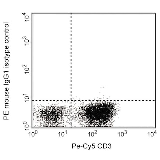
Flow cytometric analysis of CD231 (TALLA-1) expression on MOLT-4 cells. Cells from the human MOLT-4 (T lymphoblastic leukemia, ATCC CRL-1582) cell line were stained with PE Mouse IgG1, κ Isotype Control (Cat. No. 554680; dashed line histogram) or PE Mouse Anti-Human CD231 antibody (Cat. No. 566578; solid line histogram) at 1 μg/test. The fluorescence histogram showing CD231 (TALLA-1) expression (or Isotype control staining) was derived from gated events with the forward and side light-scatter characteristics of viable MOLT-4 cells. Flow cytometric analysis was performed using a BD LSRFortessa™ X-20 Flow Cytometer System. Data shown on this Technical Data Sheet are not lot specific.
.png)

BD Pharmingen™ PE Mouse Anti-Human CD231 (TALLA-1)
.png)
규제 상태 범례
Becton, Dickinson and Company의 명시적인 서면 승인 없이는 사용 하실 수 없습니다.
준비 및 보관
제품 고시
- Since applications vary, each investigator should titrate the reagent to obtain optimal results.
- An isotype control should be used at the same concentration as the antibody of interest.
- Caution: Sodium azide yields highly toxic hydrazoic acid under acidic conditions. Dilute azide compounds in running water before discarding to avoid accumulation of potentially explosive deposits in plumbing.
- For fluorochrome spectra and suitable instrument settings, please refer to our Multicolor Flow Cytometry web page at www.bdbiosciences.com/colors.
- Please refer to www.bdbiosciences.com/us/s/resources for technical protocols.
관련 제품


The M3-3D9 (SN1a) monoclonal antibody specifically recognizes CD231 which is also known as T-cell acute lymphoblastic leukemia-associated antigen 1 (TALLA-1) or Membrane component chromosome X surface marker 1 (MXS1). CD231 (TALLA-1) consists of four transmembrane domains, a short extracellular loop, a short intracellular loop, a longer extracellular loop, and short N- and C-terminal cytoplasmic tails. This ~150 kDa multi-pass transmembrane glycoprotein is encoded by TSPAN7 (Tetraspanin 7). It belongs to the tetraspanin superfamily (TM4SF) and is likewise known as Transmembrane 4 superfamily member 2 (TM4SF2). CD231 (TALLA-1) may play roles in integrin-mediated migration and the control of neurite outgrowth. It is expressed on the surface of T-cell type acute lymphoblastic leukemia (T-ALL) cells, neuroblastoma cells, and normal brain neurons. CD231 (TALLA-1) gene mutations are associated with X-linked mental retardation.

개발 참고 자료 (8)
-
Abidi FE, Holinski-Feder E, Rittinger O, et al. A novel 2 bp deletion in the TM4SF2 gene is associated with MRX58. J Med Genet. 2002; 39(6):430-433. (Biology). 참조 보기
-
Azorsa DO, Horejsi V. CD231 (TALLA-1) Summary and Workshop report. In: Mason D. David Mason .. et al., ed. Leucocyte typing VII : white cell differentiation antigens : proceedings of the Seventh International Workshop and Conference held in Harrogate, United Kingdom. Oxford: Oxford University Press; 202:509-510.
-
Bassani S, Cingolani LA, Valnegri P, et al. The X-linked intellectual disability protein TSPAN7 regulates excitatory synapse development and AMPAR trafficking. Neuron. 2012; 73(6):1143-1158. (Biology). 참조 보기
-
Castellví-Bel S, Milà M. Genes responsible for nonspecific mental retardation.. Mol Genet Metab. 2001; 72(2):104-8. (Biology). 참조 보기
-
Cheong CM, Chow AW, Fitter S, et al. Tetraspanin 7 (TSPAN7) expression is upregulated in multiple myeloma patients and inhibits myeloma tumour development in vivo. Exp Cell Res. 2015; 332(1):24-38. (Biology). 참조 보기
-
Matsuzaki H, Seon BK. Molecular nature of a cell membrane antigen specific for human T-cell acute lymphoblastic leukemia. Cancer Res. 1987; 47(16):4283-4286. (Immunogen: Blocking, Radioimmunoassay). 참조 보기
-
McLaughlin KA, Richardson CC, Ravishankar A, et al. Identification of Tetraspanin-7 as a Target of Autoantibodies in Type 1 Diabetes. Diabetes. 2016; 65(6):1690-8. (Biology). 참조 보기
-
Seon BK, Negoro S, Barcos MP. Monoclonal antibody that defines a unique human T-cell leukemia antigen.. Proc Natl Acad Sci USA. 1983; 80(3):845-9. (Biology). 참조 보기
Please refer to Support Documents for Quality Certificates
Global - Refer to manufacturer's instructions for use and related User Manuals and Technical data sheets before using this products as described
Comparisons, where applicable, are made against older BD Technology, manual methods or are general performance claims. Comparisons are not made against non-BD technologies, unless otherwise noted.
For Research Use Only. Not for use in diagnostic or therapeutic procedures.
Report a Site Issue
This form is intended to help us improve our website experience. For other support, please visit our Contact Us page.