

준비 및 보관
권장 분석 절차
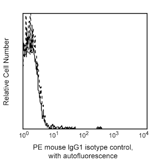
Immunofluorescent Staining and Flow Cytometric Analysis: The PE Mouse Anti-Human CD126 (Cat. No. 551850) can be used for the immunofluorescent staining (20 µg/10e6 cells) and flow cytometric analysis of human nucleated cells to measure their expressed levels of surface IL-6Ra.15 An appropriate purified immunoglobulin is PE Mouse IgG1, κ Isotype Control (Cat. No. 555749).
ELISA: Purified Mouse Anti-Human CD126 (Cat. No. 551462) is useful as a capture for a sandwich ELISA that measures soluble human IL-6Ra protein levels. The M5 antibody can be paired with the M182 antibody and recombinant soluble human IL-6Ra as a standard.
제품 고시
- This reagent has been pre-diluted for use at the recommended Volume per Test. We typically use 1 × 10^6 cells in a 100-µl experimental sample (a test).
- An isotype control should be used at the same concentration as the antibody of interest.
- Caution: Sodium azide yields highly toxic hydrazoic acid under acidic conditions. Dilute azide compounds in running water before discarding to avoid accumulation of potentially explosive deposits in plumbing.
- Ficoll-Paque is a trademark of Amersham Biosciences Limited.
- Source of all serum proteins is from USDA inspected abattoirs located in the United States.
- For fluorochrome spectra and suitable instrument settings, please refer to our Multicolor Flow Cytometry web page at www.bdbiosciences.com/colors.
- Please refer to www.bdbiosciences.com/us/s/resources for technical protocols.
관련 제품


The M5 monoclonal antibody specifically binds to human CD126 which is also known as the alpha subunit of the human IL-6 Receptor (IL-6Rα). CD126 is an 80 kDa type I transmembrane glycoprotein, also known as gp80 and B cell stimulatory factor-2 (BSF-2) Receptor. The IL-6Rα subunit associates with the 130-160 kDa gp130 subunit (IL-6 Receptor β chain, CD130), that is shared with the receptor complexes for Leukemia Inhibitory Factor (LIF), Ciliary Neurotropic Factor (CNTF), Oncostatin M (OSM), IL-11, Cardiotropin 1 (CT-1) and possibly Neurotrophin-1/B Cell-Stimulating Factor 3 (NNT-1/BSF-3). The IL-6Rα chain binds IL-6 with low affinity; however the association with CD130 stabilizes the IL-6/IL-6Rα complex resulting in the formation of a high affinity ligand-receptor complex. The IL-6Rβ chain mediates signal transduction. CD126 is expressed at high levels by activated and EBV-transformed B cells, plasma cells and myeloma cells and at lower levels by most leucocytes, epithelial cells, fibroblasts, hepatocytes and neural cells. IL-6Rα exists in soluble form in human serum. The serum levels of soluble IL-6Rα appear to elevate in pathological situations such as multiple myeloma, Grave's disease, juvenile chronic arthritis and HIV. The M5 antibody is directed against an epitope not involved in interactions of CD126 with IL-6 or CD130.

개발 참고 자료 (17)
-
Browning JL, Dougas I, Ngam-ek A, et al. Characterization of surface lymphotoxin forms. Use of specific monoclonal antibodies and soluble receptors.. J Immunol. 1995; 154(1):33-46. (Biology). 참조 보기
-
Gaillard JP, Bataille R, Brailly H, et al. Increased and highly stable levels of functional soluble interleukin-6 receptor in sera of patients with monoclonal gammopathy. Eur J Immunol. 1993 April; 23(4):820-824. (Biology). 참조 보기
-
Gaillard JP, Mani JC, Liautard J, Klein B, Brochier J. Interleukin-6 receptor signaling. I. gp80 and gp130 receptor interaction in the absence of interleukin-6.. Eur Cytokine Netw. 1999; 10(1):43-8. (Biology). 참조 보기
-
Hibi M, Murakami M, Saito M, Hirano T, Taga T, Kishimoto T. Molecular cloning and expression of an IL-6 signal transducer, gp130. Cell. 1990; 63(6):1149-1157. (Biology). 참조 보기
-
Hirano T, Nakajima K, Hibi M. Signaling mechanisms through gp130: a model of the cytokine system. Cytokine Growth Factor Rev. 1997 December; 8(4):241-252. (Biology). 참조 보기
-
Honda M, Yamamoto S, Cheng M, et al. Human soluble IL-6 receptor: its detection and enhanced release by HIV infection. J Immunol. 1992 April; 148(7):2175-2180. (Biology). 참조 보기
-
Keul R, Heinrich PC, Müller-newen G, Muller K, Woo P. A possible role for soluble IL-6 receptor in the pathogenesis of systemic onset juvenile chronic arthritis. Cytokine. 1998 September; 10(9):729-734. (Biology). 참조 보기
-
Liautard J, Gaillard JP, Mani JC, et al. Epitope analysis of human IL-6 receptor gp80 molecule with monoclonal antibodies.. Eur Cytokine Netw. 1994 May-June; 5(3):293-300. (Biology). 참조 보기
-
Llinas L, Lazaro A, de Salort J, Matesanz-Isabel J, Sintes J, Engel P. Expression profiles of novel cell surface molecules on B-cell subsets and plasma cells as analyzed by flow cytometry. Immunol Lett. 2011; 134(2):113-121. (Biology). 참조 보기
-
Müller-Newen G, Köhne C, Keul R, et al. Purification and characterization of the soluble interleukin-6 receptor from human plasma and identification of an isoform generated through alternative splicing. Eur J Biochem. 1996 March; 236(3):837-842. (Biology). 참조 보기
-
Salvi M, Girasole G, Pedrazzoni M, et al. Increased serum concentrations of interleukin-6 (IL-6) and soluble IL-6 receptor in patients with Graves' disease. J Clin Immunol. 1996 August; 81(8):2976-2979. (Biology). 참조 보기
-
Senaldi G, Varnum BC, Sarmiento U, et al. Novel neurotrophin-1/B cell-stimulating factor-3: a cytokine of the IL-6 family. Proc Natl Acad Sci U S A. 1999 September; 96(20):11458-11463. (Biology). 참조 보기
-
Taga T, Hibi M, Hirata Y, et al. Interleukin-6 triggers the association of its receptor with a possible signal transducer, gp130. Cell. 1989 August; 58(3):573-581. (Biology). 참조 보기
-
Taga T, Kawanishi Y, Hardy RR, Hirano T, Kishimoto T. Receptors for B cell stimulatory factor 2. Quantitation, specificity, distribution, and regulation of their expression. J Exp Med. 1987 October; 166(4):967-981. (Biology). 참조 보기
-
Van Snick J. Interleukin-6: an overview. Annu Rev Immunol. 1990; 8:253-278. (Biology). 참조 보기
-
Yamasaki K, Taga T, Hirata Y, et al. Cloning and expression of the human interleukin-6 (BSF-2/IFN beta 2) receptor. Science. 1988 August; 241(4867):825-828. (Biology). 참조 보기
-
Zola H. Detection of receptors for cytokines and growth factors. Immunologist. 1994; 2:47-50. (Clone-specific: Flow cytometry).
Please refer to Support Documents for Quality Certificates
Global - Refer to manufacturer's instructions for use and related User Manuals and Technical data sheets before using this products as described
Comparisons, where applicable, are made against older BD Technology, manual methods or are general performance claims. Comparisons are not made against non-BD technologies, unless otherwise noted.
For Research Use Only. Not for use in diagnostic or therapeutic procedures.