![FITC Rat Anti-Mouse Vα 3.2[b,c] TCR](/content/dam/bdb/products/global/reagents/flow-cytometry-reagents/research-reagents/single-color-antibodies-ruo/553xxx/5532xx/553219_base/01454C_553219_image1.png)
준비 및 보관
제품 고시
- Since applications vary, each investigator should titrate the reagent to obtain optimal results.
- Please refer to www.bdbiosciences.com/us/s/resources for technical protocols.
- Caution: Sodium azide yields highly toxic hydrazoic acid under acidic conditions. Dilute azide compounds in running water before discarding to avoid accumulation of potentially explosive deposits in plumbing.
관련 제품
.png?imwidth=320)
.png?imwidth=320)
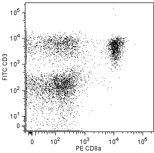
The RR3-16 monoclonal antibody specifically reacts with the Vα 3.2 T-Cell Receptor (TCR) of mice having the b (e.g., C57BL) and c (e.g., SWR, SJL, NZB, NOD) haplotypes of the Tcra gene complex, but not with TCR encoded by other members of Tcra-V3 gene subfamily. RR3-16 antibody does not react with strains having the a (e.g., A, AKR, BALB/c, CBA, C3H/He) or d (e.g., DBA/1, DBA/2, NZW) Tcra haplotypes. In addition, it has been shown that frequencies of Vα 3.2+ CD8+ T cells from homozygous H-2k/H-2k mice are moderately higher than those from heterozygous H-2k/H-2d mice, suggesting positive selection by H-2k.
This antibody is routinely tested by flow cytometric analysis. Other applications were tested at BD Biosciences Pharmingen during antibody development only or reported in the literature.

개발 참고 자료 (3)
-
Tomonari K, Fairchild S, Rosenwasser OA. Influence of viral superantigens on V beta- and V alpha-specific positive and negative selection. Immunol Rev. 1993; 131:131-168. (Biology). 참조 보기
-
Utsunomiya Y, Bill J, Palmer E, Gollob K, Takagaki Y, Kanagawa O. Analysis of a monoclonal rat antibody directed to the alpha-chain variable region (V alpha 3) of the mouse T cell antigen receptor. J Immunol. 1989; 143(8):2602-2608. (Immunogen). 참조 보기
-
Utsunomiya Y, Bill J, Palmer E, Kanagawa O. Identification of a mouse T-cell antigen receptor alpha-chain polymorphism by a V alpha 3.2 chain-specific monoclonal antibody. Immunogenetics. 1991; 33(3):198-201. (Clone-specific). 참조 보기
Please refer to Support Documents for Quality Certificates
Global - Refer to manufacturer's instructions for use and related User Manuals and Technical data sheets before using this products as described
Comparisons, where applicable, are made against older BD Technology, manual methods or are general performance claims. Comparisons are not made against non-BD technologies, unless otherwise noted.
For Research Use Only. Not for use in diagnostic or therapeutic procedures.