





.png)


준비 및 보관


제품 고시
- Since applications vary, each investigator should titrate the reagent to obtain optimal results.
- Caution: Sodium azide yields highly toxic hydrazoic acid under acidic conditions. Dilute azide compounds in running water before discarding to avoid accumulation of potentially explosive deposits in plumbing.
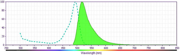
- For fluorochrome spectra and suitable instrument settings, please refer to our Multicolor Flow Cytometry web page at www.bdbiosciences.com/colors.
- Please refer to www.bdbiosciences.com/us/s/resources for technical protocols.
- An isotype control should be used at the same concentration as the antibody of interest.
.png?imwidth=320)
The GK1.5 monoclonal antibody specifically binds to the mouse CD4 (L3T4) differentiation antigen. CD4 is expressed on most thymocytes, a subpopulation of mature T lymphocytes (i.e., MHC class II-restricted T cells, including most T helper cells), and a subset of NK-T cells. In addition, CD4 has also been reported to be detectable on pluripotent hematopoietic stem cells, bone marrow myeloid and B-lymphocyte precursors, intrathymic lymphoid precursors, and a subset of splenic dendritic cells. CD4 has also been reported to be expressed on the plasma membrane of mouse egg cells and is involved in adhesion of the egg to MHC class II-bearing sperm. CD4 is an antigen coreceptor on the T-cell surface which interacts with MHC class II molecules on antigen-presenting cells. It participates in T-cell activation through its association with the T-cell receptor complex and protein tyrosine kinase lck. The GK1.5 antibody reportedly blocks binding of the RM4-5 and H129.19, but not RM4-4 mouse CD4-specific antibodies.
개발 참고 자료 (7)
-
Dialynas DP, Quan ZS, Wall KA, et al. Characterization of the murine T cell surface molecule, designated L3T4, identified by monoclonal antibody GK1.5: similarity of L3T4 to the human Leu-3/T4 molecule. J Immunol. 1983; 131(5):2445-2451. (Immunogen: Blocking, Depletion, Immunoprecipitation). 참조 보기
-
Dialynas DP, Wilde DB, Marrack P, et al. Characterization of the murine antigenic determinant, designated L3T4a, recognized by monoclonal antibody GK1.5: expression of L3T4a by functional T cell clones appears to correlate primarily with class II MHC antigen-reactivity. Immunol Rev. 1983; 74:29-56. (Clone-specific: Blocking, Depletion, Immunoprecipitation). 참조 보기
-
Frederickson GG, Basch RS. L3T4 antigen expression by hemopoietic precursor cells. J Exp Med. 1989; 169(4):1473-1478. (Biology). 참조 보기
-
Ghobrial RR, Boublik M, Winn HJ, Auchincloss H Jr. In vivo use of monoclonal antibodies against murine T cell antigens. Clin Immunol Immunopathol. 1989; 52(3):486-506. (Biology). 참조 보기
-
Guo MW, Watanabe T, Mori E, Mori T. Molecular structure and function of CD4 on murine egg plasma membrane. Zygote. 1995; 3(1):65-73. (Biology: Blocking). 참조 보기
-
Janeway CA Jr. The T cell receptor as a multicomponent signalling machine: CD4/CD8 coreceptors and CD45 in T cell activation. Annu Rev Immunol. 1992; 10:645-674. (Biology). 참조 보기
-
Wineman JP, Gilmore GL, Gritzmacher C, Torbett BE, Muller-Sieburg CE. CD4 is expressed on murine pluripotent hematopoietic stem cells. Blood. 1992; 180(7):1717-1724. (Biology). 참조 보기
Please refer to Support Documents for Quality Certificates
Global - Refer to manufacturer's instructions for use and related User Manuals and Technical data sheets before using this products as described
Comparisons, where applicable, are made against older BD Technology, manual methods or are general performance claims. Comparisons are not made against non-BD technologies, unless otherwise noted.
For Research Use Only. Not for use in diagnostic or therapeutic procedures.
