-
Reagents
- Flow Cytometry Reagents
-
Western Blotting and Molecular Reagents
- Immunoassay Reagents
-
Single-Cell Multiomics Reagents
- BD® AbSeq Assay
- BD Rhapsody™ Accessory Kits
- BD® Single-Cell Multiplexing Kit
- BD Rhapsody™ Targeted mRNA Kits
- BD Rhapsody™ Whole Transcriptome Analysis (WTA) Amplification Kit
- BD Rhapsody™ TCR/BCR Profiling Assays
- BD OMICS-Guard Sample Preservation Buffer
- BD Rhapsody™ ATAC-Seq Assays
- BD Rhapsody™ TCR/BCR Next Multiomic Assays
-
Functional Assays
-
Microscopy and Imaging Reagents
-
Cell Preparation and Separation Reagents
-
- BD® AbSeq Assay
- BD Rhapsody™ Accessory Kits
- BD® Single-Cell Multiplexing Kit
- BD Rhapsody™ Targeted mRNA Kits
- BD Rhapsody™ Whole Transcriptome Analysis (WTA) Amplification Kit
- BD Rhapsody™ TCR/BCR Profiling Assays
- BD OMICS-Guard Sample Preservation Buffer
- BD Rhapsody™ ATAC-Seq Assays
- BD Rhapsody™ TCR/BCR Next Multiomic Assays
- Korea (Korea)
- 국가 / 언어 변경
Old Browser
Looks like you're visiting us from {countryName}.
Would you like to stay on the current country site or be switched to your country?
Multiparameter flow cytometric analysis using BD OptiBuild™ BV711 Mouse Anti-Human CD217 (IL-17RA) antibody (Cat. No. 747939; Right Plot) on Human peripheral blood, with corresponding IgG Isotype Control (Cat. No. 563044; Left Plot). Flow cytometry was performed using a BD LSRFortessa™ X-20 Flow Cytometer System.
BD OptiBuild™ BV711 Mouse Anti-Human CD217 (IL-17RA)
규제 상태 범례
Becton, Dickinson and Company의 명시적인 서면 승인 없이는 사용 하실 수 없습니다.
준비 및 보관
권장 분석 절차
BD® CompBeads can be used as surrogates to assess fluorescence spillover (compensation). When fluorochrome conjugated antibodies are bound to BD® CompBeads, they have spectral properties very similar to cells. However, for some fluorochromes there can be small differences in spectral emissions compared to cells, resulting in spillover values that differ when compared to biological controls. It is strongly recommended that when using a reagent for the first time, users compare the spillover on cells and BD® CompBeads to ensure that BD® CompBeads are appropriate for your specific cellular application.
For optimal and reproducible results, BD Horizon Brilliant Stain Buffer should be used anytime BD Horizon Brilliant dyes are used in a multicolor flow cytometry panel. Fluorescent dye interactions may cause staining artifacts which may affect data interpretation. The BD Horizon Brilliant Stain Buffer was designed to minimize these interactions. When BD Horizon Brilliant Stain Buffer is used in the multicolor panel, it should also be used in the corresponding compensation controls for all dyes to achieve the most accurate compensation. For the most accurate compensation, compensation controls created with either cells or beads should be exposed to BD Horizon Brilliant Stain Buffer for the same length of time as the corresponding multicolor panel. More information can be found in the Technical Data Sheet of the BD Horizon Brilliant Stain Buffer (Cat. No. 563794/566349) or the BD Horizon Brilliant Stain Buffer Plus (Cat. No. 566385).
제품 고시
- The production process underwent stringent testing and validation to assure that it generates a high-quality conjugate with consistent performance and specific binding activity. However, verification testing has not been performed on all conjugate lots.
- Please refer to www.bdbiosciences.com/us/s/resources for technical protocols.
- Since applications vary, each investigator should titrate the reagent to obtain optimal results.
- An isotype control should be used at the same concentration as the antibody of interest.
- Caution: Sodium azide yields highly toxic hydrazoic acid under acidic conditions. Dilute azide compounds in running water before discarding to avoid accumulation of potentially explosive deposits in plumbing.
- For fluorochrome spectra and suitable instrument settings, please refer to our Multicolor Flow Cytometry web page at www.bdbiosciences.com/colors.
- Human donor specific background has been observed in relation to the presence of anti-polyethylene glycol (PEG) antibodies, developed as a result of certain vaccines containing PEG, including some COVID-19 vaccines. We recommend use of BD Horizon Brilliant™ Stain Buffer in your experiments to help mitigate potential background. For more information visit https://www.bdbiosciences.com/en-us/support/product-notices.
- Please refer to http://regdocs.bd.com to access safety data sheets (SDS).
- For U.S. patents that may apply, see bd.com/patents.
관련 제품
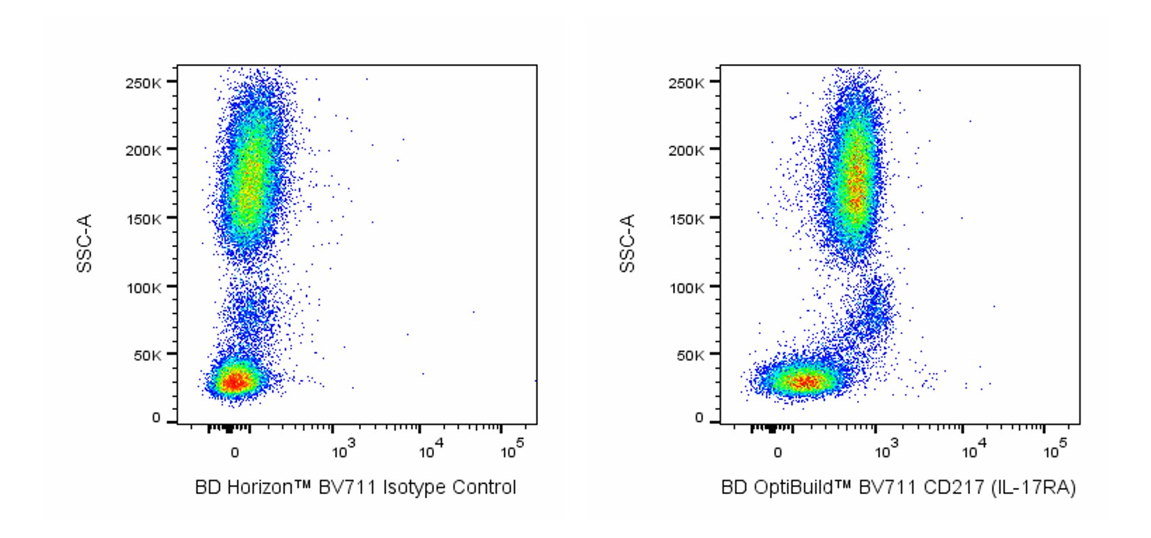
The 133617 monoclonal antibody specifically recognizes CD217 which is also known as Interleukin-17 receptor A (IL-17R or IL-17RA). CD217 is a ~120 kDa single-pass type I transmembrane glycoprotein that is encoded by IL17RA. CD217 (IL-17RA) belongs to the IL-17 receptor family which also includes IL-17 RB, IL-17RC, IL-17RD, and IL-17RE. CD217 (IL-17RA) has a wide tissue distribution and is variably expressed on granulocytes, monocytes, macrophages, dendritic cells (DCs), T cells, B cells, NK cells, smooth muscle cells, endothelial cells, and epithelial cells. Interleukin-17A (IL-17A) or IL-17F homodimers, or IL-17A:IL-17F heterodimers can bind to and signal through functional IL-17 receptor complexes comprised of CD217 (IL-17RA) and IL-17RC subunits. This leads to the cellular release of proinflammatory cytokines including IL-1, IL-6, and TNF, antimicrobial peptides, and chemokines that attract neutrophils. IL-17E/IL-25 binds to and signals through another functional IL-17 receptor complex comprised of CD217 (IL-17RA) and IL-17RB subunits. IL-17E/IL-25 induces type-2 (Th2-like) immune responses and is implicated in asthma. IL-17C binds to IL-17 receptor complexes comprised of CD217 (IL-17RA) and IL-17RE subunits and can stimulate the production of proinflammatory cytokines and antimicrobial peptides.
개발 참고 자료 (5)
-
Ahn SH, Edwards AK, Singh SS, Young SL, Lessey BA, Tayade C. IL-17A Contributes to the Pathogenesis of Endometriosis by Triggering Proinflammatory Cytokines and Angiogenic Growth Factors.. J Immunol. 2015; 195(6):2591-600. (Clone-specific: Flow cytometry). 참조 보기
-
Brucklacher-Waldert V, Steinbach K, Lioznov M, Kolster M, Hölscher C, Tolosa E. Phenotypical characterization of human Th17 cells unambiguously identified by surface IL-17A expression.. J Immunol. 2009; 183(9):5494-501. (Clone-specific: Flow cytometry). 참조 보기
-
Gaffen SL. Structure and signalling in the IL-17 receptor family.. Nat Rev Immunol. 2009; 9(8):556-67. (Biology). 참조 보기
-
Yao Z, Spriggs MK, Derry JM, et al. Molecular characterization of the human interleukin (IL)-17 receptor. Cytokine. 1997; 9(11):794-800. (Biology). 참조 보기
-
Zola H, Swart B, Banham A, et al. CD molecules 2006--human cell differentiation molecules.. J Immunol Methods. 2007; 319(1-2):1-5. (Clone-specific: Flow cytometry). 참조 보기
Please refer to Support Documents for Quality Certificates
Global - Refer to manufacturer's instructions for use and related User Manuals and Technical data sheets before using this products as described
Comparisons, where applicable, are made against older BD Technology, manual methods or are general performance claims. Comparisons are not made against non-BD technologies, unless otherwise noted.
For Research Use Only. Not for use in diagnostic or therapeutic procedures.
Report a Site Issue
This form is intended to help us improve our website experience. For other support, please visit our Contact Us page.