
준비 및 보관
권장 분석 절차
BD® CompBeads can be used as surrogates to assess fluorescence spillover (compensation). When fluorochrome conjugated antibodies are bound to BD® CompBeads, they have spectral properties very similar to cells. However, for some fluorochromes there can be small differences in spectral emissions compared to cells, resulting in spillover values that differ when compared to biological controls. It is strongly recommended that when using a reagent for the first time, users compare the spillover on cells and BD® CompBeads to ensure that BD® CompBeads are appropriate for your specific cellular application.
For optimal and reproducible results, BD Horizon Brilliant™ Stain Buffer should be used anytime BD Horizon Brilliant dyes are used in a multicolor flow cytometry panel. Fluorescent dye interactions may cause staining artifacts which may affect data interpretation. The BD Horizon Brilliant Stain Buffer was designed to minimize these interactions. When BD Horizon Brilliant Stain Buffer is used in in the multicolor panel, it should also be used in the corresponding compensation controls for all dyes to achieve the most accurate compensation. For the most accurate compensation, compensation controls created with either cells or beads should be exposed to BD Horizon Brilliant Stain Buffer for the same length of time as the corresponding multicolor panel. More information can be found in the Technical Data Sheet of the BD Horizon Brilliant Stain Buffer (Cat. No. 563794/566349) or the BD Horizon Brilliant Stain Buffer Plus (Cat. No. 566385).
제품 고시
- Please refer to www.bdbiosciences.com/us/s/resources for technical protocols.
- Since applications vary, each investigator should titrate the reagent to obtain optimal results.
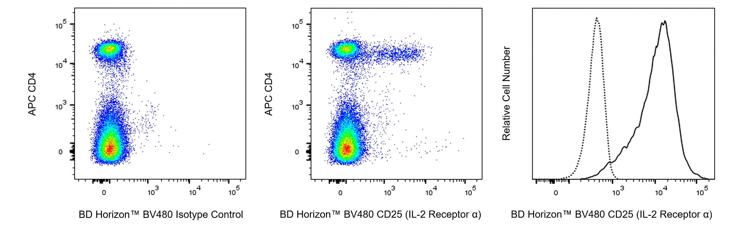
- An isotype control should be used at the same concentration as the antibody of interest.
- Caution: Sodium azide yields highly toxic hydrazoic acid under acidic conditions. Dilute azide compounds in running water before discarding to avoid accumulation of potentially explosive deposits in plumbing.
- BD Horizon Brilliant Violet 480 is covered by one or more of the following US patents: 8,575,303; 8,354,239.
- For fluorochrome spectra and suitable instrument settings, please refer to our Multicolor Flow Cytometry web page at www.bdbiosciences.com/colors.
- BD Horizon Brilliant Stain Buffer is covered by one or more of the following US patents: 8,110,673; 8,158,444; 8,575,303; 8,354,239.
- Please refer to http://regdocs.bd.com to access safety data sheets (SDS).
- For U.S. patents that may apply, see bd.com/patents.
관련 제품


The 3C7 monoclonal antibody specifically binds to CD25, the low affinity IL-2 Receptor (IL-2Rα, p55) expressed on activated T and B lymphocytes from all mouse strains tested. IL-2Rα by itself is not a signaling receptor. However, it can combine with IL-2 Receptor β (CD122) and γc (CD132) chains to form high-affinity signaling receptor complexes for IL-2. Resting T and B lymphocytes as well as resting and activated NK cells do not express IL-2Rα. CD25 is transiently expressed at a low level during normal B-cell development in the bone marrow during the CD45R/B220low TdT- sIg- Pre-B/Pre-B-II and CD45R/B220low TdT- sIgM+ sIgD- immature B stages, but not during the CD45R/B220low TdT+ sIg- Pro-B/Pre B-I stage nor on CD45R/B220high TdTsIgM+ sIgD+ mature B cells. It is expressed at a higher level during a very early stage of T-cell development in fetal and adult thymus. Peripheral CD25+ CD4+ T lymphocytes called regulatory T (Treg) cells are involved in the maintenance of self-tolerance. It has also been reported that dendritic cells express CD25 (recognized by mAb 7D4, another CD25-specific antibody). The 3C7 antibody recognizes an epitope of CD25 which is distinct from those recognized by the other CD25-specific mAbs, 7D4 and PC61. 3C7 blocks the binding of IL-2 to CD25.

개발 참고 자료 (12)
-
Chen J, Ma A, Young F, Alt FW. IL-2 receptor alpha chain expression during early B lymphocyte differentiation. Int Immunol. 1994; 6(8):1265-1268. (Clone-specific: Flow cytometry). 참조 보기
-
Crowley M, Inaba K, Witmer-Pack M, Steinman RM. The cell surface of mouse dendritic cells: FACS analyses of dendritic cells from different tissues including thymus. Cell Immunol. 1989; 118(1):108-125. (Biology). 참조 보기
-
Garni-Wagner BA, Witte PL, Tutt MM, et al. Natural killer cells in the thymus. Studies in mice with severe combined immune deficiency. J Immunol. 1990; 144(3):796-803. (Biology). 참조 보기
-
Habu S, Okumura K, Diamantstein T, Shevach EM. Expression of interleukin 2 receptor on murine fetal thymocytes. Eur J Immunol. 1985; 15(5):456-460. (Clone-specific: Flow cytometry). 참조 보기
-
Malek TR, Robb RJ, Shevach EM. Identification and initial characterization of a rat monoclonal antibody reactive with the murine interleukin 2 receptor-ligand complex. Proc Natl Acad Sci U S A. 1983; 80(18):5694-5698. (Biology). 참조 보기
-
Malek TR. The biology of interleukin-2. Annu Rev Immunol. 2008; 26:453-479. (Biology). 참조 보기
-
Moreau JL, Nabholz M, Diamantstein T, Malek T, Shevach E, Theze J. Monoclonal antibodies identify three epitope clusters on the mouse p55 subunit of the interleukin 2 receptor: relationship to the interleukin 2-binding site. Eur J Immunol. 1987; 17(7):929-935. (Clone-specific: Bioassay, Blocking, Functional assay, Inhibition, Neutralization, Radioimmunoassay). 참조 보기
-
Ortega G, Robb RJ, Shevach EM, Malek TR. The murine IL 2 receptor. I. Monoclonal antibodies that define distinct functional epitopes on activated T cells and react with activated B cells. J Immunol. 1984; 133(4):1970-1975. (Immunogen: Blocking, Flow cytometry, Immunoprecipitation, Inhibition, Neutralization, Radioimmunoassay). 참조 보기
-
Pollard AM, Lipscomb MF. Characterization of murine lung dendritic cells: similarities to Langerhans cells and thymic dendritic cells. J Exp Med. 1990; 172(1):159-167. (Biology). 참조 보기
-
Read S, Malmstrom V, Powrie F. Cytotoxic T lymphocyte-associated antigen 4 plays an essential role in the function of CD25(+)CD4(+) regulatory cells that control intestinal inflammation. J Exp Med. 2000; 192(2):295-302. (Biology). 참조 보기
-
Rolink A, Grawunder U, Winkler TH, Karasuyama H, Melchers F. IL-2 receptor alpha chain (CD25, TAC) expression defines a crucial stage in pre-B cell development. Int Immunol. 1994; 6(8):1257-1264. (Biology). 참조 보기
-
Takahashi T, Tagami T, Yamazaki S, et al. Immunologic self-tolerance maintained by CD25(+)CD4(+) regulatory T cells constitutively expressing cytotoxic T lymphocyte-associated antigen 4. J Exp Med. 2000; 192(2):303-309. (Biology). 참조 보기
Please refer to Support Documents for Quality Certificates
Global - Refer to manufacturer's instructions for use and related User Manuals and Technical data sheets before using this products as described
Comparisons, where applicable, are made against older BD Technology, manual methods or are general performance claims. Comparisons are not made against non-BD technologies, unless otherwise noted.
For Research Use Only. Not for use in diagnostic or therapeutic procedures.