







준비 및 보관


권장 분석 절차
BD™ CompBeads can be used as surrogates to assess fluorescence spillover (Compensation). When fluorochrome conjugated antibodies are bound to BD CompBeads, they have spectral properties very similar to cells. However, for some fluorochromes there can be small differences in spectral emissions compared to cells, resulting in spillover values that differ when compared to biological controls. It is strongly recommended that when using a reagent for the first time, users compare the spillover on cells and BD CompBead to ensure that BD CompBeads are appropriate for your specific cellular application.
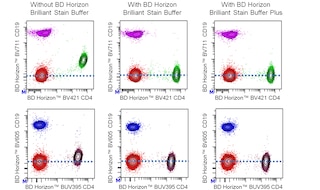
For optimal and reproducible results, BD Horizon Brilliant Stain Buffer should be used anytime two or more BD Horizon Brilliant dyes are used in the same experiment. Fluorescent dye interactions may cause staining artifacts which may affect data interpretation. The BD Horizon Brilliant Stain Buffer was designed to minimize these interactions. More information can be found in the Technical Data Sheet of the BD Horizon Brilliant Stain Buffer (Cat. No. 563794/566349) or the BD Horizon Brilliant Stain Buffer Plus (Cat. No. 566385).
Note: When using high concentrations of antibody, background binding of this dye to erythroid cell subsets (mature erythrocytes and precursors) has been observed. For researchers studying these cell populations, or in cases where light scatter gating does not adequately exclude these cells from the analysis, this background may be an important factor to consider when selecting reagents for panel(s).
제품 고시
- The production process underwent stringent testing and validation to assure that it generates a high-quality conjugate with consistent performance and specific binding activity. However, verification testing has not been performed on all conjugate lots.
- Researchers should determine the optimal concentration of this reagent for their individual applications.
- An isotype control should be used at the same concentration as the antibody of interest.
- Caution: Sodium azide yields highly toxic hydrazoic acid under acidic conditions. Dilute azide compounds in running water before discarding to avoid accumulation of potentially explosive deposits in plumbing.
- For fluorochrome spectra and suitable instrument settings, please refer to our Multicolor Flow Cytometry web page at www.bdbiosciences.com/colors.
- Please refer to www.bdbiosciences.com/us/s/resources for technical protocols.
- BD Horizon Brilliant Stain Buffer is covered by one or more of the following US patents: 8,110,673; 8,158,444; 8,575,303; 8,354,239.
- Please refer to http://regdocs.bd.com to access safety data sheets (SDS).
- CF™ is a trademark of Biotium, Inc.
- BD Horizon Brilliant Ultraviolet 615 is covered by one or more of the following US patents: 8,110,673; 8,158,444; 8,227,187; 8,575,303; 8,354,239.
관련 제품
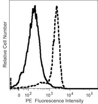



The TUGh4 monoclonal antibody specifically binds to CD132. CD132 is a 65-70 kDa type 1 transmembrane glycoprotein that is encoded by the IL2RG (interleukin 2 receptor, gamma) gene. CD132 is also known as the common γ subunit (γc) and is shared by the IL-2, IL-4, IL-7, IL-9, IL-15 and IL-21 receptor complexes. CD132 is broadly expressed by most peripheral T and B lymphocytes, NK cells, monocytes, and granulocytes. The cytoplasmic domain of the γc chain plays an important role in cytokine-mediated signal transduction. Mutation of the IL2RG gene results in X-linked severe combined immunodeficiency (XSCID). The TUGh4 antibody recognizes a different epitope from that recognized by the CD132-specific clone, AG184.
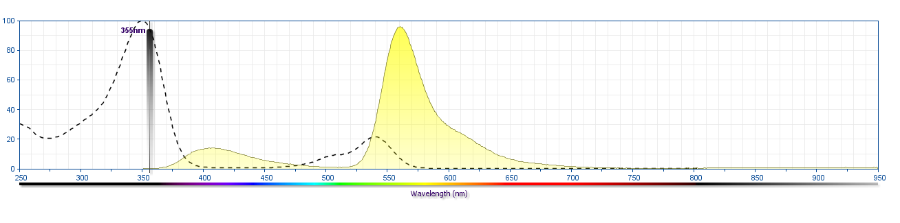
The antibody was conjugated to BD Horizon BUV615 which is part of the BD Horizon Brilliant™ Ultraviolet family of dyes. This dye is a tandem fluorochrome with an Ex Max near 350 nm and an Em Max near 615 nm. BD Horizon Brilliant BUV615 can be excited by the ultraviolet laser (355 nm) and detected with a 610/20 filter and a 595 nm LP. Due to the excitation of the acceptor dye by the blue/yellow-green laser line, there may be significant spillover into channels detecting PE-CF594 like emissions (eg, 610/20-nm filter).
개발 참고 자료 (4)
-
Ishii N, Kondo M, Takeshita T, and Sugamura K. mAb specific for the γ chain of the IL-2 receptor. In: Schlossman SF. Stuart F. Schlossman .. et al., ed. Leucocyte typing V : white cell differentiation antigens : proceedings of the fifth international workshop and conference held in Boston, USA, 3-7 November, 1993. Oxford: Oxford University Press; 1995:1867-1868.
-
Ishii N, Takeshita T, Kimura Y, et al. Expression of the IL-2 receptor gamma chain on various populations in human peripheral blood. Int Immunol. 1994; 6(8):1273-1277. (Immunogen: Flow cytometry, Immunoprecipitation, Radioimmunoassay). 참조 보기
-
Matsuoka M, Takeshita T, Ishii N, Nakamura M, Ohkubo T, Sugamura K. Kinetic study of interleukin-2 binding on the reconstituted interleukin-2 receptor complexes including the human gamma chain. Eur J Immunol. 1993; 23(10):2472-2476. (Biology). 참조 보기
-
Tanaka N, Sugamura K. CD132 (interleukin 2 γ chain (common γ) Workshop Panel report. In: Kishimoto T. Tadamitsu Kishimoto .. et al., ed. Leucocyte typing VI : white cell differentiation antigens : proceedings of the sixth international workshop and conference held in Kobe, Japan, 10-14 November 1996. New York: Garland Pub.; 1997:861-864.
Please refer to Support Documents for Quality Certificates
Global - Refer to manufacturer's instructions for use and related User Manuals and Technical data sheets before using this products as described
Comparisons, where applicable, are made against older BD Technology, manual methods or are general performance claims. Comparisons are not made against non-BD technologies, unless otherwise noted.
For Research Use Only. Not for use in diagnostic or therapeutic procedures.
