-
Reagents
- Flow Cytometry Reagents
-
Western Blotting and Molecular Reagents
- Immunoassay Reagents
-
Single-Cell Multiomics Reagents
- BD® AbSeq Assay
- BD Rhapsody™ Accessory Kits
- BD® Single-Cell Multiplexing Kit
- BD Rhapsody™ Targeted mRNA Kits
- BD Rhapsody™ Whole Transcriptome Analysis (WTA) Amplification Kit
- BD Rhapsody™ TCR/BCR Profiling Assays
- BD OMICS-Guard Sample Preservation Buffer
- BD Rhapsody™ ATAC-Seq Assays
- BD Rhapsody™ TCR/BCR Next Multiomic Assays
-
Functional Assays
-
Microscopy and Imaging Reagents
-
Cell Preparation and Separation Reagents
-
- BD® AbSeq Assay
- BD Rhapsody™ Accessory Kits
- BD® Single-Cell Multiplexing Kit
- BD Rhapsody™ Targeted mRNA Kits
- BD Rhapsody™ Whole Transcriptome Analysis (WTA) Amplification Kit
- BD Rhapsody™ TCR/BCR Profiling Assays
- BD OMICS-Guard Sample Preservation Buffer
- BD Rhapsody™ ATAC-Seq Assays
- BD Rhapsody™ TCR/BCR Next Multiomic Assays
- Korea (Korea)
- 국가 / 언어 변경
Old Browser
Looks like you're visiting us from {countryName}.
Would you like to stay on the current country site or be switched to your country?
Flow cytometric analysis of CD127 expression on human peripheral blood lymphocytes. Human whole blood was stained with either BD Horizon™ APC-R700 Mouse IgG1, κ Isotype Control (Cat. No. 564974; dashed line histogram) or BD Horizon APC-R700 Mouse Anti-Human CD127 antibody (Cat. No. 565185; solid line histogram). The erythrocytes were lysed with BD FACS™ Lysing Solution (Cat. No. 349202). The fluorescence histograms were derived from events with the forward and side light-scatter characteristics of intact lymphocytes. Flow cytometric analysis was performed using a BD™ LSR II Flow Cytometer System.
BD Horizon™ APC-R700 Mouse Anti-Human CD127
규제 상태 범례
Becton, Dickinson and Company의 명시적인 서면 승인 없이는 사용 하실 수 없습니다.
준비 및 보관
제품 고시
- This reagent has been pre-diluted for use at the recommended Volume per Test. We typically use 1 × 10^6 cells in a 100-µl experimental sample (a test).
- Please refer to www.bdbiosciences.com/us/s/resources for technical protocols.
- Alexa Fluor® is a registered trademark of Molecular Probes, Inc., Eugene, OR.
- Source of all serum proteins is from USDA inspected abattoirs located in the United States.
- Caution: Sodium azide yields highly toxic hydrazoic acid under acidic conditions. Dilute azide compounds in running water before discarding to avoid accumulation of potentially explosive deposits in plumbing.
- For fluorochrome spectra and suitable instrument settings, please refer to our Multicolor Flow Cytometry web page at www.bdbiosciences.com/colors.
- An isotype control should be used at the same concentration as the antibody of interest.
관련 제품
The hIL-7R-M21 monoclonal antibody specifically binds to the 60-90 kDa glycoprotein, CD127. CD127 is also known as the IL-7 receptor alpha (IL-7Rα) subunit. The IL-7 receptor complex is a heterodimer composed of CD127 and the common gamma chain (γc, CD132), shared by other cytokine receptors (IL-2R, IL-4R, IL-9R, IL-15R, and IL-21R). CD127 is expressed on thymocytes, T- and B-cell progenitors, mature T cells, and some lymphoid and myeloid cells. In vitro experiments show the expression of CD127 is down-regulated following T cell activation. Studies indicate that the IL-7 Receptor plays an important role in the proliferation and differentiation of mature T cells. Recently, it has been shown that low surface expression of CD127, in combination with intermediate to high surface expression of CD25, the α chain of the IL-2 receptor complex, can distinguish between human regulatory and conventional CD4+ T cells in human adult and cord blood, lymph nodes and thymus.
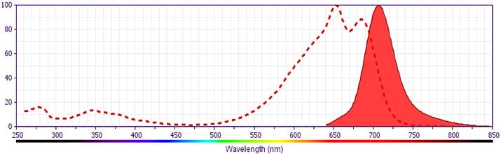
This antibody was conjugated to BD Horizon APC-R700, which has been developed exclusively by BD Biosciences as a better alternative to Alexa Fluor® 700. APC-R700 excites and emits at similar wavelengths to Alexa Fluor® 700 yet exhibits significantly improved brightness. This dye can be excited by the red laser and detected with the same filter set as Alexa Fluor® (eg, 730/45-nm filter).
개발 참고 자료 (7)
-
Appasamy PM. Biological and clinical implications of interleukin-7 and lymphopoiesis. Cytokines Cell Mol Ther. 1999; 5(1):25-39. (Biology: Flow cytometry). 참조 보기
-
Armitage RJ, Ziegler SF, Friend DJ, Park LS, Fanslow WC. Identification of a novel low-affinity receptor for human interleukin-7. Blood. 1992; 79(7):1738-1745. (Immunogen: Flow cytometry). 참조 보기
-
Benjamin D, Sharma V, Knobloch TJ, Armitage RJ, Dayton MA, Goodwin RG. B cell IL-7. Human B cell lines constitutively secrete IL-7 and express IL-7 receptors. J Immunol. 1994; 152(10):4749-4757. (Clone-specific: Flow cytometry). 참조 보기
-
Goodwin RG, Friend D, Ziegler SF et al. Cloning of the human and murine interleukin-7 receptors: demonstration of a soluble form and homology to a new receptor superfamily. Cell. 1990; 60(6):941-951. (Biology). 참조 보기
-
Hofmeister R, Khaled AR, Benbernou N, Rajnavolgyi E, Muegge K, Durum SK. Interleukin-7: physiological roles and mechanisms of action. Cytokine Growth Factor Rev. 1999; 10(1):41-60. (Biology). 참조 보기
-
Liu W, Putnam AL, Xu-Yu Z, et al. CD127 expression inversely correlates with FoxP3 and suppressive function of human CD4+ T reg cells. J Exp Med. 2006; 203(7):1701-1711. (Clone-specific: Flow cytometry, Fluorescence activated cell sorting). 참조 보기
-
Seddiki N, Santner-Nanan B, Martinson J et al. Expression of interleukin (IL)-2 and IL-7 receptors discriminates between human regulatory and activated T cells. J Exp Med. 2006; 203(7):1693-1700. (Biology). 참조 보기
Please refer to Support Documents for Quality Certificates
Global - Refer to manufacturer's instructions for use and related User Manuals and Technical data sheets before using this products as described
Comparisons, where applicable, are made against older BD Technology, manual methods or are general performance claims. Comparisons are not made against non-BD technologies, unless otherwise noted.
For Research Use Only. Not for use in diagnostic or therapeutic procedures.
Report a Site Issue
This form is intended to help us improve our website experience. For other support, please visit our Contact Us page.