Preparation And Storage
Contamination prevention: Upon first opening, prepare single-use aliquots using aseptic technique in a biosafety cabinet. Perform all subsequent handling in a biosafety cabinet to avoid contamination from repeated container entry.Long-Term Storage of Preserved Samples:• – 80°C for up to 5 months• Liquid nitrogen (Below –120°C) for up to 9 monthsPre‑cryopreservation HoldIf immediate freezing is not possible, cell suspensions may first be held in BD® OMICS‑Guard Sample Preservation Buffer at 4°C for up to 72 hours. Transfer to the CRYO buffer via a simple buffer exchange before proceeding to freezing.
Recommended Assay Procedures
Contains Bovine Serum Albumin (BSA).
Follow standard laboratory safety protocols. Review the Safety Data Sheets (SDSs) before handling chemicals.
Warning: Treat all biological specimens and materials contacting speciments as biohazardous. Dispose of waste in
accordance with applicable with federal, state, and local regulations.
Never pipette by mouth. Wear appropriate personal protective equipment, including lab coat, safety eyewear, and gloves.
I. Single-cell suspension
Recommended usage: For optimal performance, resuspend 1 × 10^5 to 2 × 10^7 cells per mL of BD® OMICS-Guard™ CRYO
Preservation Buffer.
Cryopreservation Protocol
1. Pellet single cells or dissociated cells by centrifuging at 400 × g for 5 minutes.
2. Carefully aspirate and discard the supernatant.
3. Resuspend the cells in BD® OMICS-Guard™ Cryopreservation Buffer at a density of 1 × 10^5 to 2 × 10^7 cells per mL
4. Transfer the suspension to cryopreservation tubes. Place tubes in a pre-cooled (2–8 °C) controlled-rate freezing container
e.g., Thermo Fisher Scientific Mr. Frosty™ Container (Cat# 5100-0001), prepared according to the manufacturer's instructions.
5. Place the freezing container in a –80 °C freezer overnight. Samples may remain at –80 °C for up to 5 months, or be transferred
to liquid nitrogen (below –120 °C) for storage of up to 9 months.
Thawing Protocol
Media Preparation: Complete RPMI media: RPMI1640 supplemented with 10% FBS (v/v).
1. Pre-warm the complete media to 37 °C and hold at temperature until use.
2. Remove the cryovial from storage and thaw rapidly in a 37 °C water bath for 1–2 min, or until only small ice crystals remain.
3. Add 1 mL of warm complete media to the 1 mL cell suspension (2 mL total). Gently pipette-mix five times.
4. Transfer the 2 mL cell suspension dropwise into a 15-mL conical tube containing 8 mL of warm complete media.
Mix by gently inverting the tube.
5. Centrifuge at 400 × g for 5 minutes.
6. Aspirate the supernatant without disturbing the cell pellet.
7. Either resuspend in 1 mL of warm complete media for an additional wash (see steps 8–10 below) or resuspend directly
in the buffer required for downstream applications.
Note: Steps 8–10 (second wash) may be omitted when cell input is limited, though this may result in residual debris.
8. Centrifuge at 400 x g for 5 minutes.
9. Aspirate the supernatant without disturbing the pellet.
10. Resuspend the cell pellet in the appropriate buffer for downstream applications.
II. Whole blood freezing
Recommended usage: Mix whole blood (collected with EDTA) with BD® OMICS-Guard Cryopreservation Buffer in a 1:3 ratio (v/v).
Important: Granulocytes do not survive freeze-thaw when stored in BD® OMICS-Guard Cryopreservation Buffer.
Cryopreservation protocol
1. Collect whole blood using EDTA-anticoagulant tubes.
Note: Alternative anticoagulants may be compatible but have not been validated.
2. In a separate tube, mix 1 part blood to 3 parts BD® OMICS-Guard Cryopreservation Buffer and mix by inverting 10 times.
3. Transfer the mixture to cryopreservation tubes and place in a pre-cooled (2–8 °C) controlled-rate freezing container,
e.g., Thermo Fisher Scientific Mr. Frosty™ Container, prepared as per manufacturer's instructions.
4. Place the cell freezing container in a –80 °C freezer overnight (samples may remain at -80 °C for up to 9 days).
Transfer to liquid nitrogen (below –120 °C) for long-term storage.
Thawing protocol
Media Preparation: Complete RPMI media: RPMI1640 supplemented 10% FBS (v/v).
Additional required reagents:
• 1X BD Pharm Lyse™ Lysing Buffer – equilibrate at room temperature (RT) and hold at temperature until use.
• Phosphate-buffered saline (PBS) supplemented with 2% fetal bovine serum (FBS).
1. Pre-warm the complete media to 37 °C and hold at that temperature until use.
2. Remove the cryovial from storage and thaw rapidly in a 37 °C water bath for 1–2 min, or until only small ice crystals remain.
3. Transfer the thawed whole blood sample into a 15- or 50 mL conical tube containing a least 9 mL of complete media.
Bring to a final volume of 15 or 50 mL (matching the tube size).
4. Gently invert 5 times to mix.
5. Centrifuge at 200 × g for 10 minutes at 4 °C.
6. Aspirate the supernatant without disturbing the pellet.
7. Add 10 mL of 1X BD Pharm Lyse™ Lysing Buffer per 1 mL of original whole blood equivalent.
8. Incubate on ice for 10 minutes.
9. Centrifuge at 400 × g for 5 min at 4 °C.
10. Resuspend the pellet in PBS + 2% FBS using an appropriate volume to isolate PBMCs.
11. For optimal PBMC recovery, StemCell SepMate™ PBMC Isolation Tubes (Catalog # 85415) are recommended.
Follow the manufacturer's protocol.
III. Bulk tissue freezing
Recommended usage: Prior to freezing, section bulk tissue into small pieces (up to ~40 mg each). We recommend using glass
petri dishes for sectioning.
Cryopreservation protocol
1. Section tissue and immediately immerse pieces in BD® OMICS-Guard Cryopreservation Buffer.
Alternatively, tissue may be held in BD® OMICS-Guard at 4 °C for up to 24 hours before transferring to
BD® OMICS-Guard Cryopreservation Buffer for long-term storage.
2. Transfer to cryopreservation tubes and place in a pre-cooled (2–8 °C) controlled-rate cell freezing container
(e.g., Thermo Fisher Scientific Mr. Frosty™ Container), prepared as per manufacturer's instructions.
3. Place the freezing container in a -80 °C freezer overnight, then transfer to liquid nitrogen (below -120 °C) for long-term storage.
Thawing protocol
Media Preparation: Complete RPMI media: RPMI1640 supplemented with 10% FBS (v/v).
1. Pre-warm the thawing media to 37 °C and hold at temperature until use.
2. Remove the cryovial from storage and thaw rapidly in a 37 °C water bath for 1–2 minutes, or until only small ice crystals remain.
3. Transfer the entire contents of the cryopreservation tube into a 15 ml falcon containing 10 ml of warm complete media.
4. Centrifuge at 400 × g for 5 minutes at RT.
5. Aspirate the supernatant without disturbing the pellet.
6. Proceed to tissue dissociation using the BD Horizon™ Dri Tumor & Tissue Dissociation Reagent (TTDR) protocol.
Use glass petri dishes for the dissociation step.
Note: Other reagents and approaches may be used for tissue dissociation but have not been validated.
Protocol for CITE-Seq application
BD® OMICS-Guard™ Cryopreservation Buffer is compatible with antibody staining after sample preservation.
For CITE-seq workflows using BD® AbSeq Antibody-Oligos and/or BD® OMICS-One Protein Panels,
use of the BD® AbSeq Enhancer Kit (Cat# 570750) is strongly recommended to optimize performance.
AbSeq Enhancers may be added at the BD Fc Block™ step or as a separate incubation prior to antibody-oligo staining.
Staining using 5 OMICS-One AbSeq Panels (~150-plex):
1. Prepare the BD Fc Block™ Reagent / BD® AbSeq Enhancer master mix as follows:
Component and total volume per sample
a. Stain buffer: 12.5 uL
b. BD Pharmingen™ Human BD Fc Block™: 5 uL
c. BD® AbSeq Enhancer kit: 7.5 uL (2.5 uL each AbSeq Enhancer 1-3)
Total volume: 25 uL
2. Centrifuge the sample at 800 x g for 5 minutes. Then, remove the supernatant without disturbing the pellet.
3. Resuspend the pellet in 25 µL of the final BD Fc Block™ Reagent/ BD® AbSeq Enhancer master mix from step 1.
4. Incubate the cells at RT for 10 minutes.
5. Add BD® AbSeq Antibody-Oligo cocktails per standard AbSeq staining protocols and incubate 30–60 minutes on ice.
Refer to the BD Rhapsody™ System Single-Cell Labeling with BD® AbSeq Ab-Oligos Protocol (Doc ID: 23-24262)
for details.
Staining using 1 to 3 OMICS-One AbSeq Panels (<90-plex):
1. Prepare the BD Fc Block™ Reagent and BD® AbSeq Enhancer Kit mixture as follows:
Component and total volume per sample
a. Stain buffer: 65 uL
b. BD Pharmingen™ Human BD Fc Block™: 5 uL
c. BD® AbSeq Enhancer kit: 30 uL (10 uL each AbSeq Enhancer 1-3)
Total volume: 100 uL
2. Centrifuge the sample at 800 x g for 5 minutes. Then, remove the supernatant without disturbing the pellet.
3. Resuspend the cell pellet in 100 µL of the final BD Fc Block™ Reagent/ BD® AbSeq Enhancer master mix from step 1.
4. Incubate the cells at RT for 10 minutes.
5. Add BD® AbSeq Antibody-Oligo cocktails per standard AbSeq staining protocols
(100 µL 2X BD® AbSeq Antibody-Oligo labeling MasterMix, incubate 30–60 minutes on ice).
Refer to the BD Rhapsody™ System Single-Cell Labeling with BD® AbSeq Ab-Oligos Protocol (Doc ID: 23-24262) for details.
BD Rhapsody™ Cartridge workflow modifications
Critical: For samples preserved in BD® OMICS-Guard Cryopreservation Buffer, extend the cartridge lysis step to a minimum of 5 minutes (up to 10 minutes if specified in the assay instructions).
Cartridge loading and washing
Resuspend the final washed cell pellet in 620 µL cold Sample Buffer from the BD Rhapsody™ Enhanced Cartridge Reagent V3
(catalog number 667052). Proceed to single-cell capture.
For full procedural details, refer to:
• BD Rhapsody™ HT Xpress System Extended-Lysis Single-Cell Capture and cDNA Synthesis (Doc ID 23-24983).
• BD Rhapsody™ HT Single-Cell Analysis System Extended-Lysis Single-Cell Capture and cDNA Synthesis (Doc ID 23-24984).
On-cartridge wash procedure
Perform the following steps after cell settling (8-minute incubation at RT).
1. After loading the cells into the BD Rhapsody™ 8-Lane Cartridge, incubate the cartridge at room temperature
for 8 minutes to allow cell settling.
2. Place the cartridge on the BD Rhapsody™ HT Xpress instrument and perform the wash steps as follows:
Material to load Volume (µL) 1 lane Pipette mode
Air 380 Prime/Wash
Cold Sample Buffer 380 Prime/Wash
Air 380 Prime/Wash
Cold Sample Buffer 380 Prime/Wash
Product Notices
- Please refer to www.bdbiosciences.com/us/s/resources for technical protocols.
- Please refer to http://regdocs.bd.com to access safety data sheets (SDS).
- For U.S. patents that may apply, see bd.com/patents.
- Since applications vary, each investigator should titrate the reagent to obtain optimal results.
- Source of all serum proteins is from USDA inspected abattoirs located in the United States.
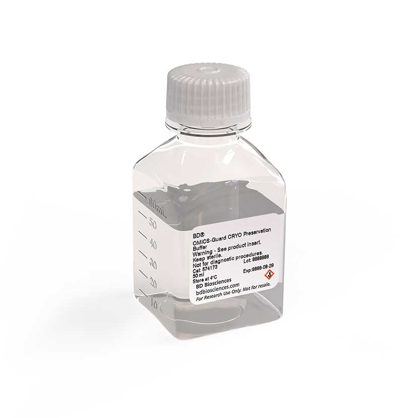
The BD® OMICS-Guard™ CRYO Preservation Buffer is a specialized cryopreservation reagent designed to maintain high-quality mRNA and protein epitopes in biological samples without cross-linking. Preserved samples are compatible with the following single-cell workflows:
• BD OMICS-One™ WTA Next Assay
• BD Rhapsody™ Multiomic ATAC-seq Assay
• BD Rhapsody™ TCR/BCR Next Assay
• BD OMICS-One™ Protein Panels
In addition, the reagent supports cell-surface protein profiling via BD® AbSeq Antibody-Oligo reagents and sample multiplexing with the BD® Single-cell Multiplexing Kit.
Important: When staining with BD® AbSeq Antibody-Oligo reagents after cryopreservation, the BD® AbSeq Enhancer must be included to obtain optimal signal recovery.
Note: For Flow Cytometry experiments, it is recommended that fluorophore‑conjugated antibody markers be evaluated individually for potential impacts on antibody binding.
No Citations Are Available for this Product
Please refer to Support Documents for Quality Certificates
Global - Refer to manufacturer's instructions for use and related User Manuals and Technical data sheets before using this products as described
Comparisons, where applicable, are made against older BD Technology, manual methods or are general performance claims. Comparisons are not made against non-BD technologies, unless otherwise noted.
For Research Use Only. Not for use in diagnostic or therapeutic procedures.