BD™ PE BCMA CAR Detection Reagent
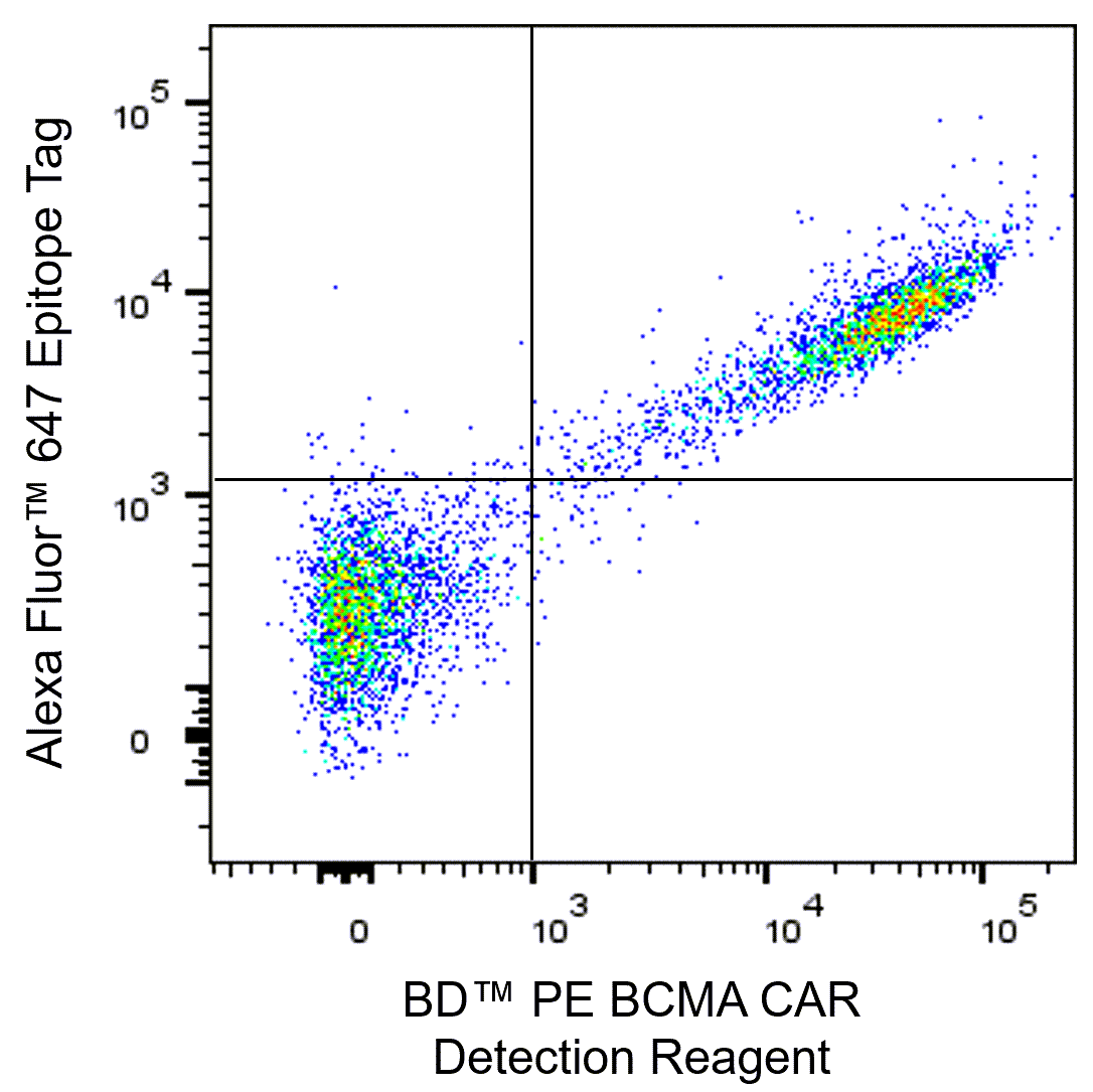
Detection of BCMA CAR expression on transfected CHO cells. CHO cells were transfected with a CAR analog construct, comprised of cell-surface Anti-Human BCMA scFv, an epitope tag, and a transmembrane domain. The cells were then stained with BD™ PE BCMA CAR Detection Reagent (Cat. No. 572803/572433) and Alexa Fluor™ 647 epitope tag-specific antibody for 30 minutes at room temperature. DAPI Solution (Cat. No. 564907) was added to cells immediately prior to analysis. The bivariate pseudocolor density plot shows BCMA CAR expression correlated with epitope tag-specific staining, gated on viable (DAPI-negative) CHO cells. Flow cytometry was performed using a BD LSRFortessa Cell Analyzer System and FlowJo™ Software.
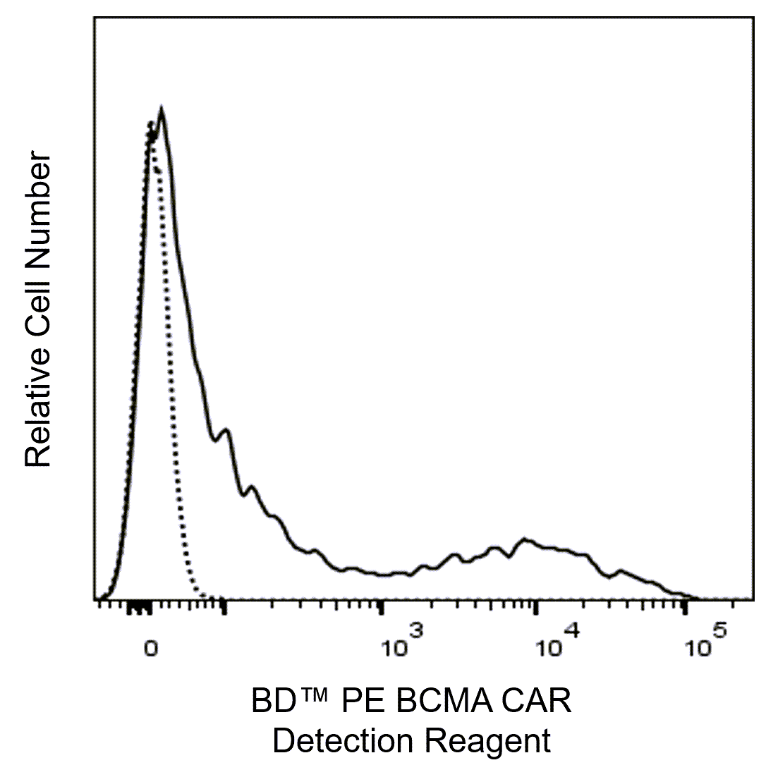
Detection of BCMA CAR expression on transduced primary Human T cells. T cells from a healthy donor were purified using the BD IMag™ Human T Lymphocyte Enrichment Set-DM (Cat. No. 557874). T cells were then activated with Anti-CD3/CD28 antibody-coupled beads plus purified BD Pharmingen™ Recombinant Human IL-2 protein (Cat. No. 554603) for 2 days, infected with BCMA CAR lentivirus, expanded for 7-10 days in culture with IL-2, and cryopreserved. Thawed BCMA CAR-transduced cells or mock transduced cells were stained with BD™ PE BCMA CAR Detection Reagent (Cat. No. 572803/572433) for 30 minutes at room temperature. DAPI solution (Cat. No. 564907) was added to cells immediately before analysis. Overlapping histograms show BCMA CAR staining with BD™ PE BCMA CAR Detection Reagent of either BCMA CAR-transduced cells (solid line) versus mock transduced cells (dashed line), gated on viable (DAPI-negative) cells. Flow cytometry was performed using a BD LSRFortessa Cell Analyzer System and FlowJo™ Software.
Detection of BCMA CAR expression on transfected CHO cells. CHO cells were transfected with a CAR analog construct, comprised of cell-surface Anti-Human BCMA scFv, an epitope tag, and a transmembrane domain. The cells were then stained with BD™ PE BCMA CAR Detection Reagent (Cat. No. 572803/572433) and Alexa Fluor™ 647 epitope tag-specific antibody for 30 minutes at room temperature. DAPI Solution (Cat. No. 564907) was added to cells immediately prior to analysis. The bivariate pseudocolor density plot shows BCMA CAR expression correlated with epitope tag-specific staining, gated on viable (DAPI-negative) CHO cells. Flow cytometry was performed using a BD LSRFortessa Cell Analyzer System and FlowJo™ Software.
Detection of BCMA CAR expression on transduced primary Human T cells. T cells from a healthy donor were purified using the BD IMag™ Human T Lymphocyte Enrichment Set-DM (Cat. No. 557874). T cells were then activated with Anti-CD3/CD28 antibody-coupled beads plus purified BD Pharmingen™ Recombinant Human IL-2 protein (Cat. No. 554603) for 2 days, infected with BCMA CAR lentivirus, expanded for 7-10 days in culture with IL-2, and cryopreserved. Thawed BCMA CAR-transduced cells or mock transduced cells were stained with BD™ PE BCMA CAR Detection Reagent (Cat. No. 572803/572433) for 30 minutes at room temperature. DAPI solution (Cat. No. 564907) was added to cells immediately before analysis. Overlapping histograms show BCMA CAR staining with BD™ PE BCMA CAR Detection Reagent of either BCMA CAR-transduced cells (solid line) versus mock transduced cells (dashed line), gated on viable (DAPI-negative) cells. Flow cytometry was performed using a BD LSRFortessa Cell Analyzer System and FlowJo™ Software.
Preparation And Storage
Recommended Assay Procedures
Surface Staining Protocol
• Aliquot 100 µl of human whole blood into tubes, or add up to 1 × 10^6 nucleated cells in 100 µl of either Stain Buffer FBS (Cat. No. 554656) or Stain Buffer BSA (Cat. No. 554657) per microwell or tube for staining.
• Add 5 μl of the BCMA CAR Detection Reagent and incubate at either 4°C or at RT for 30 minutes protected from light.
• For stained whole blood samples, treat with either BD Pharm Lyse™ Lysing Buffer (Cat. No. 555899) or with BD FACS™ Lysing Solution (Cat. No. 349202). Wash cells with stain buffer and proceed with acquisition.
• For stained PBMC or other nucleated cell samples, wash cells with stain buffer, add viability dye and proceed with acquisition.
Notes:
• Recommend using BD Pharmingen™ Human BD Fc Block™ (Cat. No. 564220) prior to staining as needed.
• This CAR Detection Reagent has been tested for compatibility with either BD Cytofix™ Fixation Buffer (Cat. No. 554655) and BD Perm/Wash™ Buffer (Cat. No. 554723) or the BD Pharmingen™ Transcription Factor Buffer Set (Cat. No. 562574).
• Avoid co-stains with Human BCMA-specific antibodies when using this reagent.
Bead-based compensation or unmixing controls, eg, BD™ SpectraComp™, can be used as surrogates to assess fluorescence spillover when bound to fluorochrome-conjugated antibodies or IgG Fc-fusion proteins. Although these beads have spectral properties similar to cells, variations in spectral emission may occur, resulting in differing spillover values compared to biological controls. Therefore, it is considered best practice to compare the spillover obtained from cells and bead-based controls when using BD™ SpectraComp™ for the first time, to ensure they are appropriate for the intended application.
Product Notices
- Please refer to www.bdbiosciences.com/us/s/resources for technical protocols.
- Source of all serum proteins is from USDA inspected abattoirs located in the United States.
- Caution: Sodium azide yields highly toxic hydrazoic acid under acidic conditions. Dilute azide compounds in running water before discarding to avoid accumulation of potentially explosive deposits in plumbing.
- This reagent has been pre-diluted for use at the recommended Volume per Test. We typically use 1 × 10^6 cells in a 100-µl experimental sample (a test).
- Please refer to http://regdocs.bd.com to access safety data sheets (SDS).
- For U.S. patents that may apply, see bd.com/patents.
- For fluorochrome spectra and suitable instrument settings, please refer to our Multicolor Flow Cytometry web page at www.bdbiosciences.com/colors.
Companion Products


Chimeric Antigen Receptors (CAR) are engineered transmembrane proteins containing an extracellular single-chain domain from a proven antibody fused with major intracellular signaling components from the T Cell Receptor (TCR) complex. A CAR thus combines a defined antibody specificity with the functional capabilities of an effector cell and is a popular approach for cellular immunotherapy of cancer or autoimmunity. In CAR T cell therapy, T cells are isolated from a patient's blood, activated, transduced with a CAR gene, expanded, and reinfused into the patient, where they proliferate upon encounter with target antigen and eliminate antigen-expressing cells. Flow cytometry is used throughout this workflow to assess CAR expression and CAR-T phenotype pre-infusion, and post-infusion to probe expansion, persistence, and in vivo CAR-T characteristics. BCMA CAR Detection Reagent is a recombinant Human BCMA (aa 1-54, extracellular domain)-Mouse IgG2a Fc fusion protein conjugated to a fluorochrome that enables 1-step staining, alone or in multicolor panels, for flow cytometric analysis of BMCA CAR-expressing T, NK, or other cells.

Development References (5)
-
Ali SA, Shi V, Maric I, et al. T cells expressing an anti-B-cell maturation antigen chimeric antigen receptor cause remissions of multiple myeloma.. Blood. 2016; 128(13):1688-700. (Biology). View Reference
-
Hansen DK, Peres LC, Dima D, et al. Comparison of Standard-of-Care Idecabtagene Vicleucel and Ciltacabtagene Autoleucel in Relapsed/Refractory Multiple Myeloma.. J Clin Oncol. 2025; 43(13):1597-1609. (Biology). View Reference
-
Hu Y, Huang J. The Chimeric Antigen Receptor Detection Toolkit.. Front Immunol. 2020; 11:1770. (Biology). View Reference
-
Scheller L, Tebuka E, Rambau PF, et al. BCMA CAR-T cells in multiple myeloma-ready for take-off?. Leuk Lymphoma. 2024; 65(2):143-157. (Biology). View Reference
-
Suarez ER, Chang de K, Sun J, et al. Chimeric antigen receptor T cells secreting anti-PD-L1 antibodies more effectively regress renal cell carcinoma in a humanized mouse model.. Oncotarget. 2016; 7(23):34341-55. (Biology). View Reference
Please refer to Support Documents for Quality Certificates
Global - Refer to manufacturer's instructions for use and related User Manuals and Technical data sheets before using this products as described
Comparisons, where applicable, are made against older BD Technology, manual methods or are general performance claims. Comparisons are not made against non-BD technologies, unless otherwise noted.
For Research Use Only. Not for use in diagnostic or therapeutic procedures.
Although not required, these products are manufactured in accordance with Good Manufacturing Practices.