





BD Horizon™ BV605 Rat Anti-Mouse CD45
Clone 30-F11 (RUO)


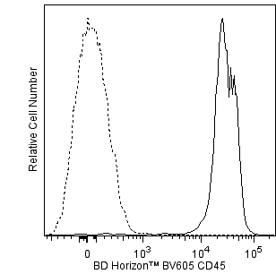
Flow cytometric analysis of CD45 expressed on mouse splenocytes. Splenic leucocytes from a BALB/c mouse were preincubated with Purified Rat Anti-Mouse CD16/CD32 antibody (Mouse BD Fc Block™) (Cat. No. 553141/553142). The cells were then stained with either BD Horizon™ BV605 Rat IgG2b, κ Isotype Control (Cat. No. 563145; dashed line histogram) or a BD Horizon™ BV605 Rat Anti-Mouse CD45 antibody (Cat.No. 563053; solid line histogram). Flow cytometric fluorescence histograms were derived from gated events based on forward and side light-scatter characteristics of viable splenocytes. Flow cytometric analysis was performed using a BD™ LSR II Flow Cytometer System.
Flow cytometric analysis of CD45 expressed on mouse splenocytes. Splenic leucocytes from a BALB/c mouse were preincubated with Purified Rat Anti-Mouse CD16/CD32 antibody (Mouse BD Fc Block™) (Cat. No. 553141/553142). The cells were then stained with either BD Horizon™ BV605 Rat IgG2b, κ Isotype Control (Cat. No. 563145; dashed line histogram) or a BD Horizon™ BV605 Rat Anti-Mouse CD45 antibody (Cat.No. 563053; solid line histogram). Flow cytometric fluorescence histograms were derived from gated events based on forward and side light-scatter characteristics of viable splenocytes. Flow cytometric analysis was performed using a BD™ LSR II Flow Cytometer System.


Preparation And Storage


Recommended Assay Procedures
For optimal and reproducible results, BD Horizon Brilliant™ Stain Buffer should be used anytime BD Horizon Brilliant™ dyes are used in a multicolor flow cytometry panel. Fluorescent dye interactions may cause staining artifacts which may affect data interpretation. The BD Horizon Brilliant Stain Buffer was designed to minimize these interactions. When BD Horizon Brilliant Stain Buffer is used in in the multicolor panel, it should also be used in the corresponding compensation controls for all dyes to achieve the most accurate compensation. For the most accurate compensation, compensation controls created with either cells or beads should be exposed to BD Horizon Brilliant Stain Buffer for the same length of time as the corresponding multicolor panel. More information can be found in the Technical Data Sheet of the BD Horizon Brilliant Stain Buffer (Cat. No. 563794/566349) or the BD Horizon Brilliant Stain Buffer Plus (Cat. No. 566385).
Product Notices
- Since applications vary, each investigator should titrate the reagent to obtain optimal results.
- An isotype control should be used at the same concentration as the antibody of interest.
- Please observe the following precautions: Absorption of visible light can significantly alter the energy transfer occurring in any tandem fluorochrome conjugate; therefore, we recommend that special precautions be taken (such as wrapping vials, tubes, or racks in aluminum foil) to prevent exposure of conjugated reagents, including cells stained with those reagents, to room illumination.
- Caution: Sodium azide yields highly toxic hydrazoic acid under acidic conditions. Dilute azide compounds in running water before discarding to avoid accumulation of potentially explosive deposits in plumbing.
- For fluorochrome spectra and suitable instrument settings, please refer to our Multicolor Flow Cytometry web page at www.bdbiosciences.com/colors.
- Although every effort is made to minimize the lot-to-lot variation in the efficiency of the fluorochrome energy transfer, differences in the residual emission from BD Horizon™ BV421 may be observed. Therefore, we recommend that individual compensation controls be performed for every BD Horizon™ BV605 conjugate.
- CF™ is a trademark of Biotium, Inc.
- BD Horizon Brilliant Violet 605 is covered by one or more of the following US patents: 8,110,673; 8,158,444; 8,227,187; 8,455,613; 8,575,303; 8,354,239.
- BD Horizon Brilliant Stain Buffer is covered by one or more of the following US patents: 8,110,673; 8,158,444; 8,575,303; 8,354,239.
- Please refer to www.bdbiosciences.com/us/s/resources for technical protocols.
Companion Products


The 30-F11 clone has been reported to react with all isoforms and both alloantigens of CD45, which is found on hematopoietic stem cells and all cells of hematopoietic origin, except erythrocytes. CD45 is a transmembrane glycoprotein which is expressed at high levels on the cell surface, and its presence distinguishes leukocytes from non-hematopoietic cells. CD45 is a member of the Protein Tyrosine Phosphatase (PTP) family, where the intracellular carboxy-terminal region contains two PTP catalytic domains, and the extracellular region is highly variable due to alternative splicing of exons 4, 5, and 6 (designated as A, B, and C, respectively). CD45 isoforms play complex roles in T-cell and B-cell antigen receptor signal transduction and the CD45 isoforms detected in the mouse are cell type-, maturation-, and activation state-specific.

Development References (7)
-
Johnson P, Maiti A, Ng DHW. CD45: A family of leukocyte-specific cell surface glycoproteins. In: Herzenberg LA, Weir DM, Herzenberg LA, Blackwell C , ed. Weir's Handbook of Experimental Immunology, Vol 2. Cambridge: Blackwell Science; 1997:62.1-62.16.
-
Lagasse E, Connors H, Al-Dhalimy M, et al. Purified hematopoietic stem cells can differentiate into hepatocytes in vivo. Nat Med. 2000; 6(11):1212-1213. (Biology). View Reference
-
Ledbetter JA, Herzenberg LA. Xenogeneic monoclonal antibodies to mouse lymphoid differentiation antigens. Immunol Rev. 1979; 47:63-90. (Immunogen: Cytotoxicity, Immunoprecipitation). View Reference
-
Simon DI, Dhen Z, Seifert P, Edelman ER, Ballantyne CM, Rogers C. Decreased neointimal formation in Mac-1(-/-) mice reveals a role for inflammation in vascular repair after angioplasty. J Clin Pathol. 2000; 105(3):293-300. (Clone-specific: Immunohistochemistry). View Reference
-
Tamaki K, Yasaka N, Chang CH, et al. Identification and characterization of novel dermal Thy-1 antigen-bearing dendritic cells in murine skin. J Invest Dermatol. 1996; 106(3):571-575. (Clone-specific: Immunofluorescence). View Reference
-
Tan J, Town T, Paris D, et al. Microglial activation resulting from CD40-CD40L interaction after beta-amyloid stimulation. Science. 1999; 286(5448):2352-2355. (Clone-specific: Immunohistochemistry). View Reference
-
Thomas ML. The leukocyte common antigen family. Annu Rev Immunol. 1989; 7:339-369. (Biology). View Reference
Please refer to Support Documents for Quality Certificates
Global - Refer to manufacturer's instructions for use and related User Manuals and Technical data sheets before using this products as described
Comparisons, where applicable, are made against older BD Technology, manual methods or are general performance claims. Comparisons are not made against non-BD technologies, unless otherwise noted.
For Research Use Only. Not for use in diagnostic or therapeutic procedures.