


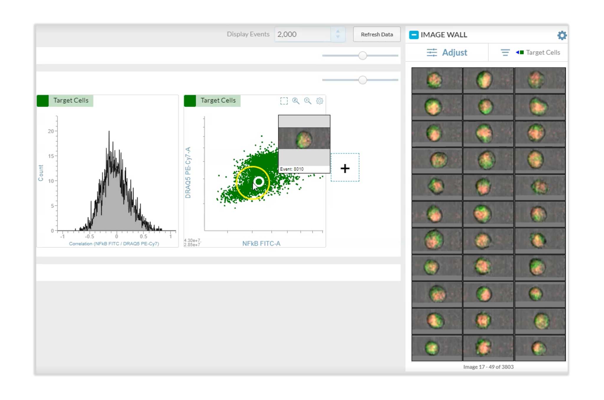
BD CellView™ Image Technology is real-time imaging (RTI) technology that combines flow cytometry data with spatial and morphological insights. The technology enables answering complex biological questions quickly, such as how cells grow, function and interact, or studying locations of viruses or proteins within a cell. The technology seamlessly integrates image and flow data on each event and can easily visualize the image for an event during acquisition. It enhances fluorescence activated cell sorting (FACS) with live visual inspection of target cells and novel gating strategies based on real-time image feature analysis using spatial distribution of fluorescence capabilities.
BD CellView™ Image Technology versus camera-based imaging cytometry
In contrast to camera-based technologies that combine imaging and flow cytometry, BD CellView™ Image Technology does not use a camera to image cells, which enables imaging at much faster rates. Camera-based technologies are limited by closely controlled fluidics and cannot be used for high-speed droplet cell sorting. BD CellView™ Image Technology implements orthogonal frequency domain multiplexing (OFDM) for imaging cells using electronic and optical components already used in flow cytometers. This method allows faster throughput than camera-based technologies. The image data can be acquired and quantified in real time at high speed for analysis and sorting as the sample is being acquired.
Principles of BD CellView™ Image Technology

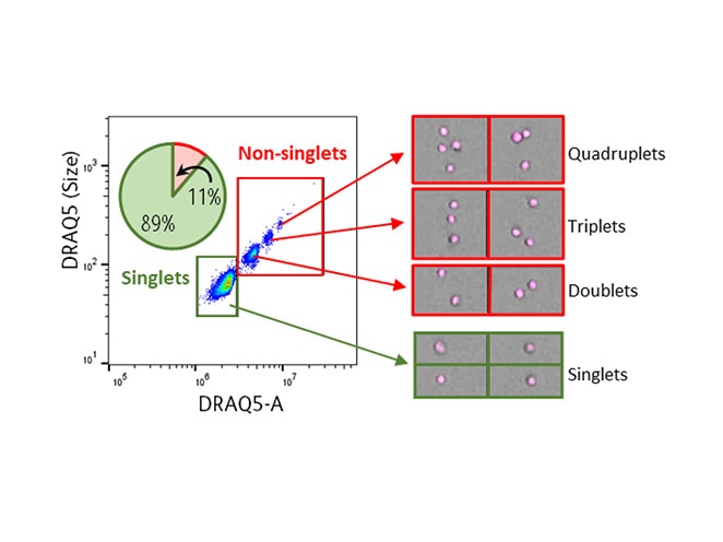
Eccentricity
Eccentricity is a ratio of the shortest to the longest axis of the identified particle (as identified by the region of analysis)
Usage examples: Doublet discrimination, cluster identification
Availability: All imaging channels

Max Intensity
Max intensity is the intensity of the brightest pixel in the image. It is not affected by the region of analysis.
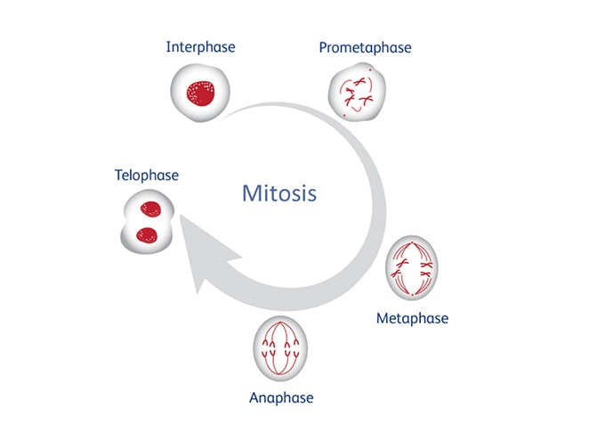
Usage examples: Punctate fluorescence, phagocytosis assay, cell cycle analysis
Availability: All imaging channels

Size
Size is the number of pixels in the image, which are brighter than a user-defined pixel threshold.
Usage examples: Label-free sorting, punctate fluorescence
Availability: All imaging channels

Radial Moment
Radial moment is the average distance of the pixels from the centroid within the region of analysis.
Usage examples: Doublet discrimination (with Eccentricity), cell—cell interactions (cellular synapse)
Availability: All imaging channels

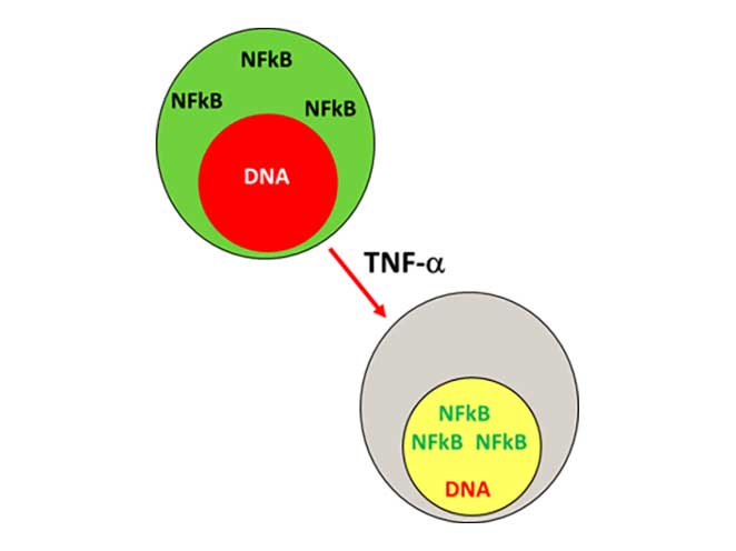
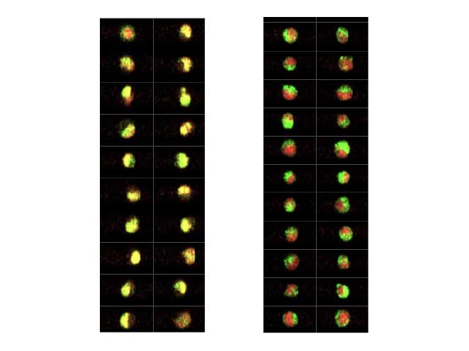
Correlation
Correlation is the degree to which the location of two imaging channels are the same within the region of pixels defined by the region of analysis.
Usage example: Translocation assay
Availability: Any two imaging fluorescence channels

Delta Center of Mass
The distance between two fluorescent signal sources in any two imaging channels within a particle as defined by the region of analysis.
Availability: Any two imaging channels

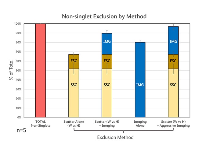
Forward Scatter (FSC)
As particles (cells) pass through the laser, the interaction of the light with the particle results in scatter in all directions.
The forward scatter detector is placed in line with the laser path to measure light that is scattered at small angles. forward scatter loosely correlates to particle (cell) size.
")
Side Scatter (SSC)
As particles (cells) pass through the laser, the interaction of the light with the particle results in scatter in all directions.
The side scatter detector measures light that is scattered perpendicular (90°) to the laser path. Side scatter loosely correlates to optical density or complexity of the particle.
")
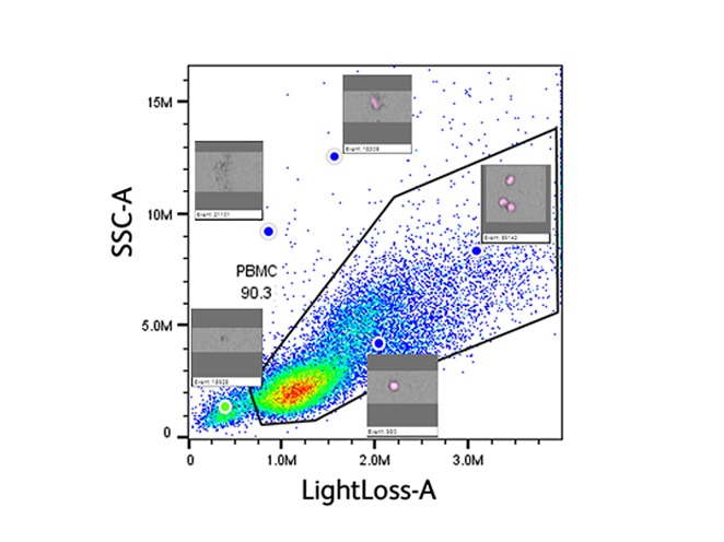
Light Loss (Imaging)
As particles (cells) pass through the laser, the interaction of the light with the particle results in scatter in all directions.
Light loss is a measure of light (photons) lost from the laser due to scattering and absorption of light by a particle (cell).
")
-DNA-Thumbnail.jpg)


Request Quote / General Inquiry
If you have questions related to a product or application, or would like to request samples, a quote or demo, please submit your inquiry.
*Required fields
For Research Use Only. Not for use in diagnostic or therapeutic procedures. CF is a trademark of Biotium, Inc.
Class 1 Laser Products.