-
Instruments
-
Flow Cytometers
- BD FACSDiscover™ Spectral Flow Cytometers Platform
-
BD FACSLyric™ Clinical Cell Analyzer
- BD FACSymphony™ Cell Analyzers & Sorters Platform
- BD LSRFortessa™ Flow Cytometer Platform
- BD FACSAria™ Cell Sorter Platform
- BD Accuri™ C6 Plus
-
BD FACSCelesta™ Cell Analyzer
-
BD FACSMelody™ Cell Sorter
-
BD FACSCanto™ Clinical Flow Cytometry System
- Clinical Sample Prep Systems
- Single-Cell Multiomics Systems
-
Flow Cytometers
-
Reagents
-
Flow Cytometry Reagents
-
Clinical Diagnostics
- ASR Single Color Antibodies
- Buffers and Supporting Reagents (IVD, CE-IVD)
- Process and Quality Controls
- Single Color Antibodies ASR IVD CE IVD
- Multicolor Cocktails and Kits IVD CE IVDs
- OneFlow
- Stem Cell Enumeration Kit Reagents
- Leucocount ™ Kit | BD Biosciences
- BD Multitest™ 6-Color TBNK Kit | BD Biosciences
- Clinical Discovery
-
Research Reagents
- Quality and Reproducibility
- Single Color Antibodies RUO
- Panels Multicolor Cocktails RUO
- Flow Cytometry Controls and Lysates
- buffers and Supporting Reagents RUO
- Cell Function Analysis Stains Dyes
- Single Color Antibodies
- Compensation Beads
- BD Horizon™ Human T Cell Backbone Panel
- BD Pharmingen™ MonoBlock™ Leukocyte Staining Buffer
- BV605 Transition
- BD Horizon Brilliant™ Dyes
- Single-Color Antibodies for Flow Cytometry (RUO)
-
BD Horizon™ Real Dyes for Flow Cytometry
- BD Horizon RealYellow™ 610
- BD Horizon RealRed™ 688 Reagents
- BD Horizon RealYellow™ 703
- BD Horizon RealBlue™ 670 for Flow Cytometry
- BD Horizon RealYellow™ 743
- BD Horizon RealBlue™ 780 for Flow Cytometry
- BD Horizon RealBlue™ 575 for Flow Cytometry | BD Real Dyes
- BD Horizon RealYellow™ 586
- BD Horizon RealBlue™ 824 for Flow Cytometry
- BD Horizon RealViolet™ 828 for Flow Cytometry
- BD Horizon RealViolet™ 544 for Flow Cytometry | BD Real Dyes
-
Clinical Diagnostics
-
Western Blotting and Molecular Reagents
- Immunoassay Reagents
-
Single-Cell Multiomics Reagents
-
BD OMICS-Guard™ CRYO Preservation Buffer
-
BD OMICS-Guard™ Sample Preservation Buffer
-
BD OMICS-One™ Protein Panels
- BD® OMICS-One Tumor Protein Panel
- BD® OMICS-One T-Cell Protein Panel
- BD® OMICS-One B-Cell Protein Panel
- BD® OMICS-One Adaptive Protein Panel
- BD® OMICS-One Immune Profiler Protein Panel
- BD® OMICS-One Immuno-Oncology Protein Panel
- BD® OMICS-One NK-Cell Protein Panel
- BD® OMICS-One APC/Myeloid-Cell Protein Panel
- BD® OMICS-One Innate Protein Panel
- BD® OMICS-One Innate Immuno-Oncology Protein Panel
- BD® OMICS-One Comprehensive Immuno-Oncology Protein Panel
- BD® AbSeq Assay
- BD® Single-Cell Multiplexing Kit
-
BD OMICS-One™ WTA Next Assay
-
BD Rhapsody™ ATAC-Seq Assays
-
BD Rhapsody™ TCR/BCR Next Multiomic Assays
-
BD Rhapsody™ Targeted mRNA Kits
-
BD Rhapsody™ Accessory Kits
-
BD OMICS-Guard™ CRYO Preservation Buffer
- Functional Assays
-
Microscopy and Imaging Reagents
-
Cell Preparation and Separation Reagents
-
Dehydrated Culture Media
-
Flow Cytometry Reagents
-
BD Biosciences Training Program
- Flow Cytometry Basic Training
-
Product-Based Training
- BD Accuri™ C6 Plus Cell Analyzer
- BD FACSAria™ Cell Sorter Cell Sorter
- BD FACSCanto™ Cell Analyzer
- BD FACSDiscover™ A8 Cell Analyzer
- BD FACSDiscover™ S8 Cell Sorter
- BD FACSDuet™ Sample Preparation System
- BD FACSLyric™ Cell Analyzer
- BD FACSMelody™ Cell Sorter
- BD FACSymphony™ Cell Analyzer
- BD LSRFortessa™ Cell Analyzer
- Advanced Training
-
-
-
-
- ASR Single Color Antibodies
- Buffers and Supporting Reagents (IVD, CE-IVD)
- Process and Quality Controls
- Single Color Antibodies ASR IVD CE IVD
- Multicolor Cocktails and Kits IVD CE IVDs
- OneFlow
- Stem Cell Enumeration Kit Reagents
- Leucocount ™ Kit | BD Biosciences
- BD Multitest™ 6-Color TBNK Kit | BD Biosciences
-
- Quality and Reproducibility
- Single Color Antibodies RUO
- Panels Multicolor Cocktails RUO
- Flow Cytometry Controls and Lysates
- buffers and Supporting Reagents RUO
- Cell Function Analysis Stains Dyes
- Single Color Antibodies
- Compensation Beads
- BD Horizon™ Human T Cell Backbone Panel
- BD Pharmingen™ MonoBlock™ Leukocyte Staining Buffer
- BV605 Transition
- BD Horizon Brilliant™ Dyes
- Single-Color Antibodies for Flow Cytometry (RUO)
-
- BD Horizon RealYellow™ 610
- BD Horizon RealRed™ 688 Reagents
- BD Horizon RealYellow™ 703
- BD Horizon RealBlue™ 670 for Flow Cytometry
- BD Horizon RealYellow™ 743
- BD Horizon RealBlue™ 780 for Flow Cytometry
- BD Horizon RealBlue™ 575 for Flow Cytometry | BD Real Dyes
- BD Horizon RealYellow™ 586
- BD Horizon RealBlue™ 824 for Flow Cytometry
- BD Horizon RealViolet™ 828 for Flow Cytometry
- BD Horizon RealViolet™ 544 for Flow Cytometry | BD Real Dyes
-
-
-
-
- BD® OMICS-One Tumor Protein Panel
- BD® OMICS-One T-Cell Protein Panel
- BD® OMICS-One B-Cell Protein Panel
- BD® OMICS-One Adaptive Protein Panel
- BD® OMICS-One Immune Profiler Protein Panel
- BD® OMICS-One Immuno-Oncology Protein Panel
- BD® OMICS-One NK-Cell Protein Panel
- BD® OMICS-One APC/Myeloid-Cell Protein Panel
- BD® OMICS-One Innate Protein Panel
- BD® OMICS-One Innate Immuno-Oncology Protein Panel
- BD® OMICS-One Comprehensive Immuno-Oncology Protein Panel
-
-
-
- United States (English)
-
Change location/language
Old Browser
This page has been recently translated and is available in French now.
Looks like you're visiting us from {countryName}.
Would you like to stay on the current location site or be switched to your location?
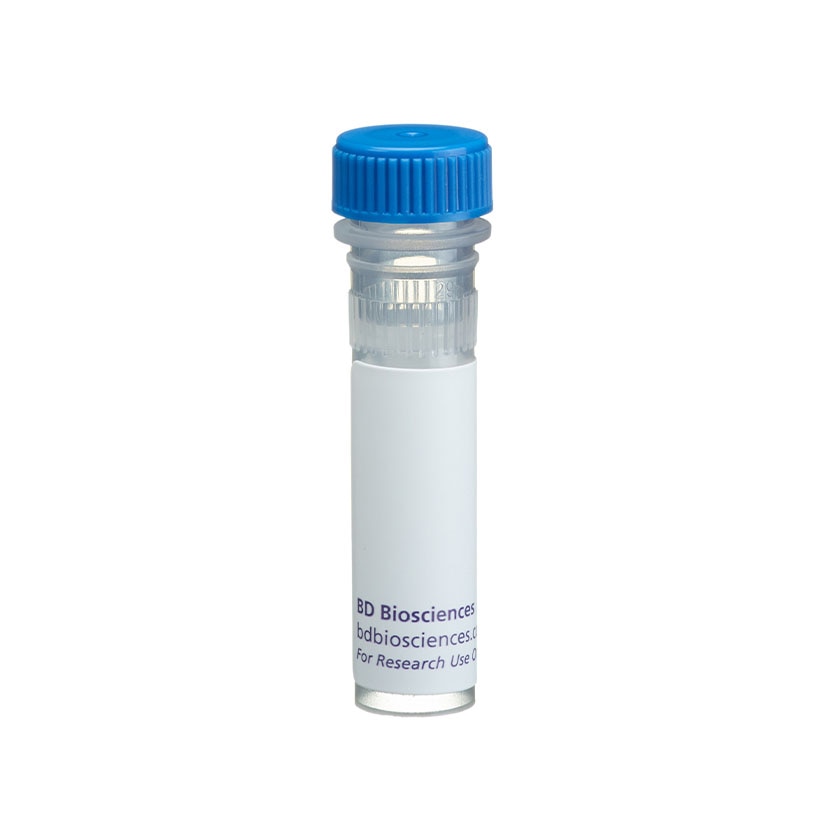
BD Transduction Laboratories™ Purified Mouse Anti-P-Cadherin
Clone 56/P-Cadherin (RUO)
Western blot analysis of P-Cadherin on A431 cell lysate. Lane 1: 1:250, lane 2: 1:500, lane 3: 1:1000 dilution of anti-P-Cadherin antibody.
Immunofluorescent staining of A431 cells.
Regulatory Status Legend
Any use of products other than the permitted use without the express written authorization of Becton, Dickinson and Company is strictly prohibited.
Preparation And Storage
Recommended Assay Procedures
Western blot: Please refer to http://www.bdbiosciences.com/pharmingen/protocols/Western_Blotting.shtml.
Product Notices
- Since applications vary, each investigator should titrate the reagent to obtain optimal results.
- Please refer to www.bdbiosciences.com/us/s/resources for technical protocols.
- Caution: Sodium azide yields highly toxic hydrazoic acid under acidic conditions. Dilute azide compounds in running water before discarding to avoid accumulation of potentially explosive deposits in plumbing.
- Source of all serum proteins is from USDA inspected abattoirs located in the United States.
Companion Products

Cadherins are a family of transmembrane glycoproteins involved in the Ca2+-dependent cell-cell adhesion that occurs in many tissues. Cadherins are similar in their domain structure, Ca2+ and protease sensitivity, and molecular weight. However, these proteins have distinct tissue expression patterns and immunological reactivities. P-Cadherin is localized in placenta while E-Cadherin and N-Cadherin are found in epithelial and neural tissues, respectively. P-Cadherin is an 829 amino acid polypeptide with a putative signal peptide and precursor region, an extracellular domain containing several internal repeats, and a highly hydrophobic transmembrane region. The cytoplasmic domain provides a link to the cytoskeleton through the associated catenin proteins.
Development References (5)
-
Bailey T, Biddlestone L, Shepherd N, Barr H, Warner P, Jankowski J. Altered cadherin and catenin complexes in the Barrett's esophagus-dysplasia-adenocarcinoma sequence: correlation with disease progression and dedifferentiation. Am J Pathol. 1998; 152(1):135-144. (Clone-specific: Immunohistochemistry). View Reference
-
Holsinger LJ, Ward K, Duffield B, Zachwieja J, Jallal B. The transmembrane receptor protein tyrosine phosphatase DEP1 interacts with p120(ctn). Oncogene. 2002; 21(46):7067-7076. (Clone-specific: Western blot). View Reference
-
Kantak SS, Kramer RH. E-cadherin regulates anchorage-independent growth and survival in oral squamous cell carcinoma cells. J Biol Chem. 1998; 273(27):16953-16961. (Clone-specific: Western blot). View Reference
-
Nose A, Nagafuchi A, Takeichi M. Isolation of placental cadherin cDNA: identification of a novel gene family of cell-cell adhesion molecules. EMBO J. 1987; 6(12):3655-3661. (Biology). View Reference
-
Tao YS, Edwards RA, Tubb B, Wang S, Bryan J, McCrea PD. beta-Catenin associates with the actin-bundling protein fascin in a noncadherin complex. J Cell Biol. 1996; 134(5):1271-1281. (Clone-specific: Immunofluorescence). View Reference
Please refer to Support Documents for Quality Certificates
Global - Refer to manufacturer's instructions for use and related User Manuals and Technical data sheets before using this products as described
Comparisons, where applicable, are made against older BD Technology, manual methods or are general performance claims. Comparisons are not made against non-BD technologies, unless otherwise noted.
For Research Use Only. Not for use in diagnostic or therapeutic procedures.