

Preparation And Storage
Product Notices
- This reagent has been pre-diluted for use at the recommended Volume per Test. We typically use 1 × 10^6 cells in a 100-µl experimental sample (a test).
- An isotype control should be used at the same concentration as the antibody of interest.
- Caution: Sodium azide yields highly toxic hydrazoic acid under acidic conditions. Dilute azide compounds in running water before discarding to avoid accumulation of potentially explosive deposits in plumbing.
- Source of all serum proteins is from USDA inspected abattoirs located in the United States.
- The Alexa Fluor®, Pacific Blue™, and Cascade Blue® dye antibody conjugates in this product are sold under license from Molecular Probes, Inc. for research use only, excluding use in combination with microarrays, or as analyte specific reagents. The Alexa Fluor® dyes (except for Alexa Fluor® 430), Pacific Blue™ dye, and Cascade Blue® dye are covered by pending and issued patents.
- Alexa Fluor® is a registered trademark of Molecular Probes, Inc., Eugene, OR.
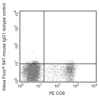
- Alexa Fluor® 647 fluorochrome emission is collected at the same instrument settings as for allophycocyanin (APC).
- For fluorochrome spectra and suitable instrument settings, please refer to our Multicolor Flow Cytometry web page at www.bdbiosciences.com/colors.
- Please refer to www.bdbiosciences.com/us/s/resources for technical protocols.
Companion Products


The R32-1149 monoclonal antibody specifically binds to Ikaros, which is encoded by IKZF1 (IKAROS family zinc finger 1). Ikaros is also known as Lymphoid transcription factor LyF-1 (LYF1) and ZNFN1A1 [Zinc finger protein, subfamily 1A, 1 (Ikaros)]. Ikaros belongs to the IKAROS family of zinc-finger transcription factors. This DNA-binding transcription factor forms homodimers and heterodimers with other IKAROS family members. It is involved in the development of lymphoid tissues and remains abundantly expressed in the thymus and by cells within peripheral lymphoid tissues. Ikaros functions in the maturation, differentiation, and homeostasis of various hematopoietic cells including T lymphocytes, B cells, NK cells, and neutrophils. Ikaros regulates the expression of a number of genes including various genes expressed during early stages of B- and T-cell development. Several alternatively-spliced human Ikaros isoforms have been described. Abnormal expression of different Ikaros isoforms has been associated with hematologic malignancies. Flow cytometric analysis of 293F cells transfected with members of the IKAROS family has demonstrated that clone R32-1149 is negative on Helios, Aiolos, and Eos transfectants.
Development References (8)
-
Billot K, Parizot C, Arrouss I, et al. Differential aiolos expression in human hematopoietic subpopulations. Leuk Res. 2010; 34(3):289-293. (Biology). View Reference
-
Billot K, Soeur J, Chereau F, et al. Deregulation of Aiolos expression in chronic lymphocytic leukemia is associated with epigenetic modifications. Blood. 2011; 117(6):1917-1927. (Biology). View Reference
-
Liippo J, Nera KP, Veistinen E, et al. Both normal and leukemic B lymphocytes express multiple isoforms of the human Aiolos gene. Eur J Immunol. 2001; 31(12):3469-3474. (Biology). View Reference
-
Morgan B, Sun L, Avitahl N, et al. Aiolos, a lymphoid restricted transcription factor that interacts with Ikaros to regulate lymphocyte differentiation. EMBO J. 1997; 16(8):2004-2013. (Biology). View Reference
-
Rebollo A, Ayllon V, Fleischer A, Martinez CA, Zaballos A. The association of Aiolos transcription factor and Bcl-xL is involved in the control of apoptosis. J Immunol. 2001; 167(11):6366-6373. (Biology). View Reference
-
Romero F, Martinez AC, Camonis J, Rebollo A. Aiolos transcription factor controls cell death in T cells by regulating Bcl-2 expression and its cellular localization. EMBO J. 1999; 18(12):3419-3430. (Biology). View Reference
-
Schmitt C, Tonnelle C, Dalloul A, Chabannon C, Debre P, Rebollo A. Aiolos and Ikaros: regulators of lymphocyte development, homeostasis and lymphoproliferation. Apoptosis. 2002; 7(3):277-284. (Biology). View Reference
-
Wang JH, Avitahl N, Cariappa A, et al. Aiolos regulates B cell activation and maturation to effector state. Immunity. 1998; 9(4):543-553. (Biology). View Reference
Please refer to Support Documents for Quality Certificates
Global - Refer to manufacturer's instructions for use and related User Manuals and Technical data sheets before using this products as described
Comparisons, where applicable, are made against older BD Technology, manual methods or are general performance claims. Comparisons are not made against non-BD technologies, unless otherwise noted.
For Research Use Only. Not for use in diagnostic or therapeutic procedures.