

Preparation And Storage
Recommended Assay Procedures
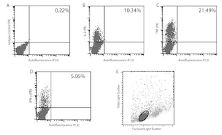
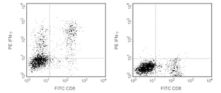
Flow cytometry: The XMG1.2 antibody is useful for immunofluorescent staining and flow cytometric analysis to identify and enumerate IFN-γ producing cells within mixed cell populations. A useful control investigators may consider using for demonstrating specificity of staining, is to pre-block with one of the following reagents: (1) recombinant mouse IFN-γ (Cat. No. 554587) or (2) unlabeled XMG1.2 antibody (Cat. No. 554409), prior to staining.
Cell Preparation: Investigators not wishing to utilize MiCK-1 cells may alternatively prepare mouse splenocytes (e.g BALB/c) stimulated for 4-6 hours with PMA (5 ng/mL, Sigma-Aldrich Cat. No. P-8139) and ionomycin (500 ng, Sigma-Aldrich Cat. No. I-0634) in the presence of 1 µg/mL Brefeldin A (BD GolgiPlug™ MN 555029). Investigators are advised to fix and permeabilize the cells prior to staining.
Product Notices
- Since applications vary, each investigator should titrate the reagent to obtain optimal results.
- An isotype control should be used at the same concentration as the antibody of interest.
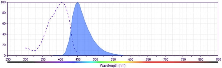
- BD Horizon V450 has a maximum absorption of 406 nm and maximum emission of 450 nm. Before staining with this reagent, please confirm that your flow cytometer is capable of exciting the fluorochrome and discriminating the resulting fluorescence.
- Caution: Sodium azide yields highly toxic hydrazoic acid under acidic conditions. Dilute azide compounds in running water before discarding to avoid accumulation of potentially explosive deposits in plumbing.
- Pacific Blue™ is a trademark of Molecular Probes, Inc., Eugene, OR.
- For fluorochrome spectra and suitable instrument settings, please refer to our Multicolor Flow Cytometry web page at www.bdbiosciences.com/colors.
- Please refer to www.bdbiosciences.com/us/s/resources for technical protocols.
Companion Products

The XMG1.2 monoclonal antibody specifically binds to mouse interferon-γ (IFN-γ) protein. IFN-γ is a pleiotropic cytokine, of approximately 15-17 kDa, involved in the regulation of inflammatory and immune responses. It plays an important role in activation, growth, and differentiation of T and B lymphocytes, macrophages, NK cells and other non-hematopoietic cell types. IFN-γ production is associated with the Th1 cell differentiation. The purified form of this antibody has been reported to be a neutralizing antibody.
The antibody is conjugated to BD Horizon™ V450, which has been developed for use in multicolor flow cytometry experiments and is available exclusively from BD Biosciences. It is excited by the Violet laser Ex max of 406 nm and has an Em Max at 450 nm. Conjugates with BD Horizon™ V450 can be used in place of Pacific Blue™ conjugates.
Development References (3)
-
Cherwinski HM, Schumacher JH, Brown KD, Mosmann TR. Two types of mouse helper T cell clone. III. Further differences in lymphokine synthesis between Th1 and Th2 clones revealed by RNA hybridization, functionally monospecific bioassays, and monoclonal antibodies. J Exp Med. 1987; 166(5):1229-1244. (Biology). View Reference
-
Prussin C, Metcalfe DD. Detection of intracytoplasmic cytokine using flow cytometry and directly conjugated anti-cytokine antibodies. J Immunol Methods. 1995; 188(1):117-128. (Methodology: Flow cytometry). View Reference
-
Sander B, Hoiden I, Andersson U, Moller E, Abrams JS. Similar frequencies and kinetics of cytokine producing cells in murine peripheral blood and spleen. Cytokine detection by immunoassay and intracellular immunostaining. J Immunol Methods. 1993; 166(2):201-214. (Methodology: Flow cytometry). View Reference
Please refer to Support Documents for Quality Certificates
Global - Refer to manufacturer's instructions for use and related User Manuals and Technical data sheets before using this products as described
Comparisons, where applicable, are made against older BD Technology, manual methods or are general performance claims. Comparisons are not made against non-BD technologies, unless otherwise noted.
For Research Use Only. Not for use in diagnostic or therapeutic procedures.