-
Instruments
-
Flow Cytometers
- BD FACSDiscover™ Spectral Flow Cytometers Platform
-
BD FACSLyric™ Clinical Cell Analyzer
- BD FACSymphony™ Cell Analyzers & Sorters Platform
- BD LSRFortessa™ Flow Cytometer Platform
- BD FACSAria™ Cell Sorter Platform
- BD Accuri™ C6 Plus
-
BD FACSCelesta™ Cell Analyzer
-
BD FACSMelody™ Cell Sorter
-
BD FACSCanto™ Clinical Flow Cytometry System
- Clinical Sample Prep Systems
- Single-Cell Multiomics Systems
-
Flow Cytometers
-
Reagents
-
Flow Cytometry Reagents
-
Clinical Diagnostics
- ASR Single Color Antibodies
- Buffers and Supporting Reagents (IVD, CE-IVD)
- Process and Quality Controls
- Single Color Antibodies ASR IVD CE IVD
- Multicolor Cocktails and Kits IVD CE IVDs
- OneFlow
- Stem Cell Enumeration Kit Reagents
- Leucocount ™ Kit | BD Biosciences
- BD Multitest™ 6-Color TBNK Kit | BD Biosciences
- Clinical Discovery
-
Research Reagents
- Quality and Reproducibility
- Single Color Antibodies RUO
- Panels Multicolor Cocktails RUO
- Flow Cytometry Controls and Lysates
- buffers and Supporting Reagents RUO
- Cell Function Analysis Stains Dyes
- Single Color Antibodies
- Compensation Beads
- BD Horizon™ Human T Cell Backbone Panel
- BD Pharmingen™ MonoBlock™ Leukocyte Staining Buffer
- BV605 Transition
- BD Horizon Brilliant™ Dyes
- Single-Color Antibodies for Flow Cytometry (RUO)
-
BD Horizon™ Real Dyes for Flow Cytometry
- BD Horizon RealYellow™ 610
- BD Horizon RealRed™ 688 Reagents
- BD Horizon RealYellow™ 703
- BD Horizon RealBlue™ 670 for Flow Cytometry
- BD Horizon RealYellow™ 743
- BD Horizon RealBlue™ 780 for Flow Cytometry
- BD Horizon RealBlue™ 575 for Flow Cytometry | BD Real Dyes
- BD Horizon RealYellow™ 586
- BD Horizon RealBlue™ 824 for Flow Cytometry
- BD Horizon RealViolet™ 828 for Flow Cytometry
- BD Horizon RealViolet™ 544 for Flow Cytometry | BD Real Dyes
-
Clinical Diagnostics
-
Western Blotting and Molecular Reagents
- Immunoassay Reagents
-
Single-Cell Multiomics Reagents
-
BD OMICS-Guard™ CRYO Preservation Buffer
-
BD OMICS-Guard™ Sample Preservation Buffer
-
BD OMICS-One™ Protein Panels
- BD® OMICS-One Tumor Protein Panel
- BD® OMICS-One T-Cell Protein Panel
- BD® OMICS-One B-Cell Protein Panel
- BD® OMICS-One Adaptive Protein Panel
- BD® OMICS-One Immune Profiler Protein Panel
- BD® OMICS-One Immuno-Oncology Protein Panel
- BD® OMICS-One NK-Cell Protein Panel
- BD® OMICS-One APC/Myeloid-Cell Protein Panel
- BD® OMICS-One Innate Protein Panel
- BD® OMICS-One Innate Immuno-Oncology Protein Panel
- BD® OMICS-One Comprehensive Immuno-Oncology Protein Panel
- BD® AbSeq Assay
- BD® Single-Cell Multiplexing Kit
-
BD OMICS-One™ WTA Next Assay
-
BD Rhapsody™ ATAC-Seq Assays
-
BD Rhapsody™ TCR/BCR Next Multiomic Assays
-
BD Rhapsody™ Targeted mRNA Kits
-
BD Rhapsody™ Accessory Kits
-
BD OMICS-Guard™ CRYO Preservation Buffer
- Functional Assays
-
Microscopy and Imaging Reagents
-
Cell Preparation and Separation Reagents
-
Dehydrated Culture Media
-
Flow Cytometry Reagents
-
BD Biosciences Training Program
- Flow Cytometry Basic Training
-
Product-Based Training
- BD Accuri™ C6 Plus Cell Analyzer
- BD FACSAria™ Cell Sorter Cell Sorter
- BD FACSCanto™ Cell Analyzer
- BD FACSDiscover™ A8 Cell Analyzer
- BD FACSDiscover™ S8 Cell Sorter
- BD FACSDuet™ Sample Preparation System
- BD FACSLyric™ Cell Analyzer
- BD FACSMelody™ Cell Sorter
- BD FACSymphony™ Cell Analyzer
- BD LSRFortessa™ Cell Analyzer
- Advanced Training
-
-
-
-
- ASR Single Color Antibodies
- Buffers and Supporting Reagents (IVD, CE-IVD)
- Process and Quality Controls
- Single Color Antibodies ASR IVD CE IVD
- Multicolor Cocktails and Kits IVD CE IVDs
- OneFlow
- Stem Cell Enumeration Kit Reagents
- Leucocount ™ Kit | BD Biosciences
- BD Multitest™ 6-Color TBNK Kit | BD Biosciences
-
- Quality and Reproducibility
- Single Color Antibodies RUO
- Panels Multicolor Cocktails RUO
- Flow Cytometry Controls and Lysates
- buffers and Supporting Reagents RUO
- Cell Function Analysis Stains Dyes
- Single Color Antibodies
- Compensation Beads
- BD Horizon™ Human T Cell Backbone Panel
- BD Pharmingen™ MonoBlock™ Leukocyte Staining Buffer
- BV605 Transition
- BD Horizon Brilliant™ Dyes
- Single-Color Antibodies for Flow Cytometry (RUO)
-
- BD Horizon RealYellow™ 610
- BD Horizon RealRed™ 688 Reagents
- BD Horizon RealYellow™ 703
- BD Horizon RealBlue™ 670 for Flow Cytometry
- BD Horizon RealYellow™ 743
- BD Horizon RealBlue™ 780 for Flow Cytometry
- BD Horizon RealBlue™ 575 for Flow Cytometry | BD Real Dyes
- BD Horizon RealYellow™ 586
- BD Horizon RealBlue™ 824 for Flow Cytometry
- BD Horizon RealViolet™ 828 for Flow Cytometry
- BD Horizon RealViolet™ 544 for Flow Cytometry | BD Real Dyes
-
-
-
-
- BD® OMICS-One Tumor Protein Panel
- BD® OMICS-One T-Cell Protein Panel
- BD® OMICS-One B-Cell Protein Panel
- BD® OMICS-One Adaptive Protein Panel
- BD® OMICS-One Immune Profiler Protein Panel
- BD® OMICS-One Immuno-Oncology Protein Panel
- BD® OMICS-One NK-Cell Protein Panel
- BD® OMICS-One APC/Myeloid-Cell Protein Panel
- BD® OMICS-One Innate Protein Panel
- BD® OMICS-One Innate Immuno-Oncology Protein Panel
- BD® OMICS-One Comprehensive Immuno-Oncology Protein Panel
-
-
-
- United States (English)
-
Change location/language
Old Browser
This page has been recently translated and is available in French now.
Looks like you're visiting us from {countryName}.
Would you like to stay on the current location site or be switched to your location?
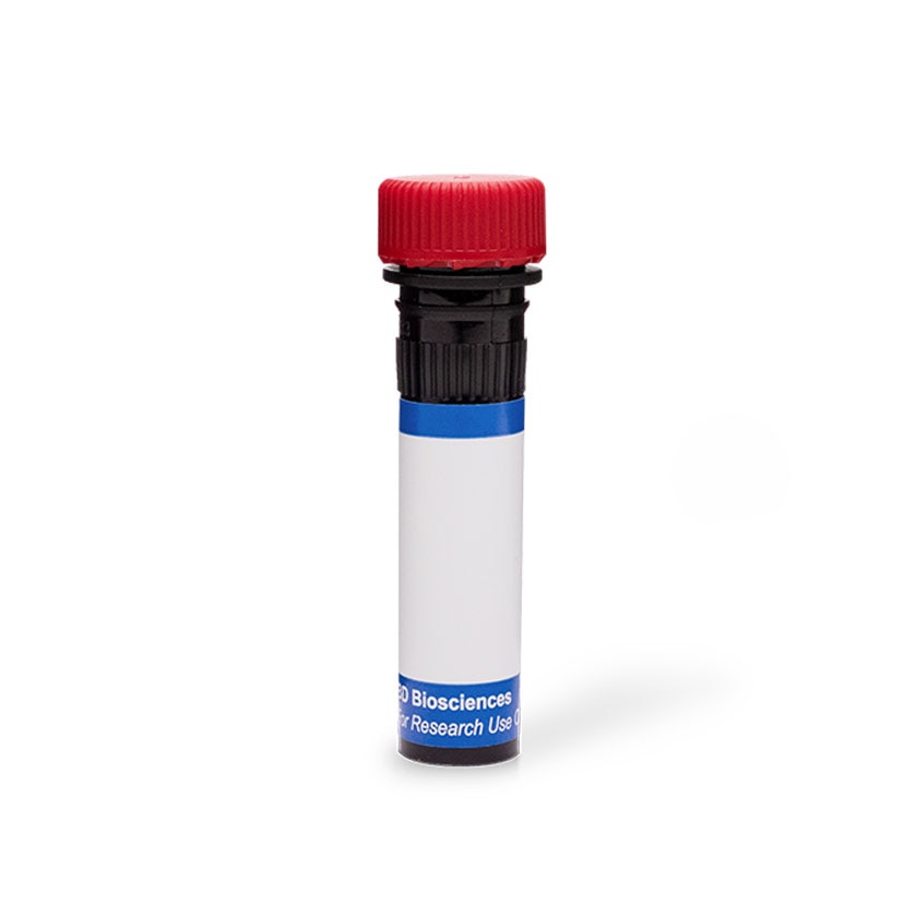
BD Pharmingen™ PE Mouse Anti-Human CD106
Clone 51-10C9 (RUO)
Flow cytometric analysis of CD106 on TNF-α-activated HUVEC cell line. HUVEC cells were stimulated with Recombinant Human TNF Protein (Cat. No. 554618; 20 ng/ml), then stained with either PE Mouse IgG1, κ Isotype Control (Cat. No.555749; dashed line histogram) or PE Mouse Anti-Human CD106 (Cat. No. 555647/561679; solid line histogram). Fluorescent histograms were derived from gated events based on the light scattering characteristics for viable HUVEC cells. Flow cytometry was performed on a BD FACScan™.
Regulatory Status Legend
Any use of products other than the permitted use without the express written authorization of Becton, Dickinson and Company is strictly prohibited.
Preparation And Storage
Product Notices
- This reagent has been pre-diluted for use at the recommended Volume per Test. We typically use 1 × 10^6 cells in a 100-µl experimental sample (a test).
- An isotype control should be used at the same concentration as the antibody of interest.
- Source of all serum proteins is from USDA inspected abattoirs located in the United States.
- Caution: Sodium azide yields highly toxic hydrazoic acid under acidic conditions. Dilute azide compounds in running water before discarding to avoid accumulation of potentially explosive deposits in plumbing.
- For fluorochrome spectra and suitable instrument settings, please refer to our Multicolor Flow Cytometry web page at www.bdbiosciences.com/colors.
- Please refer to www.bdbiosciences.com/us/s/resources for technical protocols.
Companion Products
The 51-10C9 monoclonal antibody specifically binds to CD106. CD106 is a 100-110 kDa type I transmembrane sialoglycoprotrein that is also known as Vascular cell adhesion molecule-1 (VCAM-1) and INCAM-110. CD106 is expressed at high levels on the surface of cytokine-stimulated endothelium, and at minimal levels on unstimulated endothelium. VCAM-1 serves as a ligand for the leukocyte integrins α4β1 (CD49d/CD29 complex; VLA-4) and α4β7 (LPAM-1). The 51-10C9 monoclonal antibody inhibits the in vitro binding of lymphocytes and monocytes to VCAM-1 on stimulated endothelium.
Development References (4)
-
Bevilacqua MP, Pober JS, Mendrick DL, Cotran RS, Gimbrone MA Jr. Identification of an inducible endothelial-leukocyte adhesion molecule. Proc Natl Acad Sci U S A. 1987; 84(24):9238-9242. (Biology). View Reference
-
Schlossman SF. Stuart F. Schlossman .. et al., ed. Leucocyte typing V : white cell differentiation antigens : proceedings of the fifth international workshop and conference held in Boston, USA, 3-7 November, 1993. Oxford: Oxford University Press; 1995.
-
Taichman DB, Cybulsky MI, Djaffar I, et al. Tumor cell surface alpha 4 beta 1 integrin mediates adhesion to vascular endothelium: demonstration of an interaction with the N-terminal domains of INCAM-110/VCAM-1. Cell Regul. 1991; 2(5):347-355. (Biology). View Reference
-
van Vugt MJ, van den Herik-Oudijk IE, van de Winkle JG. Binding of PE-CY5 conjugates to the human high-affinity receptor for IgG (CD64). Blood. 1996; 88(6):2358-2361. (Immunogen). View Reference
Please refer to Support Documents for Quality Certificates
Global - Refer to manufacturer's instructions for use and related User Manuals and Technical data sheets before using this products as described
Comparisons, where applicable, are made against older BD Technology, manual methods or are general performance claims. Comparisons are not made against non-BD technologies, unless otherwise noted.
For Research Use Only. Not for use in diagnostic or therapeutic procedures.