
Three-color analysis of erythroid cells in the rat bone marrow. LEW rat bone marrow cells were simultaneously stained with PE Mouse Anti-Rat CD71 (Cat. no. 554891), BD Horizon™ V450 Mouse Anti-Rat CD45 (Cat. No. 561587), and APC Mouse anti-Rat Erythroid Cells (Cat. No. 562348). The two-color dot plot of CD45 versus CD71 expression is displayed in Panel A. The remaining panels (Panels B-F) display histograms of Erythroid Cells antigen expression on the gated subpopulations shown in Panel A: Panel B, CD45+ CD71+ bright immature erythroid cells; Panel C, CD45- CD71+ maturing erythroid cells; Panel D, CD45- CD71- erythrocytes; Panel E, CD45+ CD71+ hematopoietic progenitors; and Panel F, CD45+ CD71- leukocytes. Flow cytometry was performed using a BD™ LSR II Flow Cytometer System.
Three-color analysis of erythroid cells in the rat bone marrow. LEW rat bone marrow cells were simultaneously stained with PE Mouse Anti-Rat CD71 (Cat. no. 554891), BD Horizon™ V450 Mouse Anti-Rat CD45 (Cat. No. 561587), and APC Mouse anti-Rat Erythroid Cells (Cat. No. 562348). The two-color dot plot of CD45 versus CD71 expression is displayed in Panel A. The remaining panels (Panels B-F) display histograms of Erythroid Cells antigen expression on the gated subpopulations shown in Panel A: Panel B, CD45+ CD71+ bright immature erythroid cells; Panel C, CD45- CD71+ maturing erythroid cells; Panel D, CD45- CD71- erythrocytes; Panel E, CD45+ CD71+ hematopoietic progenitors; and Panel F, CD45+ CD71- leukocytes. Flow cytometry was performed using a BD™ LSR II Flow Cytometer System.
Product Details


BD Pharmingen™
Rat (QC Testing)
Mouse BALB/c IgM, κ
Rat bone marrow cells
Flow cytometry (Routinely Tested)
0.2 mg/ml
AB_11152955
Aqueous buffered solution containing protein stabilizer and ≤0.09% sodium azide.
RUO
Preparation And Storage
Store undiluted at 4°C and protected from prolonged exposure to light. Do not freeze. The monoclonal antibody was purified from tissue culture supernatant or ascites by affinity chromatography. The antibody was conjugated to APC under optimum conditions, and unconjugated antibody and free APC were removed.
Product Notices
- Since applications vary, each investigator should titrate the reagent to obtain optimal results.
- Caution: Sodium azide yields highly toxic hydrazoic acid under acidic conditions. Dilute azide compounds in running water before discarding to avoid accumulation of potentially explosive deposits in plumbing.
- This APC-conjugated reagent can be used in any flow cytometer equipped with a dye, HeNe, or red diode laser.
- For fluorochrome spectra and suitable instrument settings, please refer to our Multicolor Flow Cytometry web page at www.bdbiosciences.com/colors.
- An isotype control should be used at the same concentration as the antibody of interest.
- Please refer to www.bdbiosciences.com/us/s/resources for technical protocols.
Companion Products
Stain Buffer (FBS) RUO
Size
500 mL
Cat No.
554656

APC Mouse IgM, κ Isotype Control RUO
Size
0.1 mg
Cat No.
550883
.png?imwidth=320)
PE Mouse Anti-Rat CD71 RUO
Size
0.2 mg
Cat No.
554891
.png?imwidth=320)
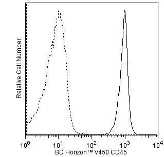
V450 Mouse Anti-Rat CD45 RUO
Size
50 µg
Cat No.
561587
562348 Rev. 1
Antibody Details
HIS49
The HIS49 antibody reacts with an antigen found on erythrocytes and erythroid progenitors, but not on granulocytes, monocytes, or lymphocytes.
562348 Rev. 1
Format Details
APC
Allophycocyanin (APC), is part of the BD family of phycobiliprotein dyes. This fluorochrome is a multimeric fluorescent phycobiliprotein with excitation maximum (Ex Max) of 651 nm and an emission maximum (Em Max) at 660 nm. APC is designed to be excited by the Red (627-640 nm) laser and detected using an optical filter centered near 660 nm (e.g., a 660/20 nm bandpass filter). Please ensure that your instrument’s configurations (lasers and optical filters) are appropriate for this dye.

APC
Red 627-640 nm
651 nm
660 nm
562348 Rev.1
Citations & References
Development References (1)
-
Hermans MH, Opstelten D. In situ visualization of hemopoietic cell subsets and stromal elements in rat and mouse bone marrow by immunostaining of frozen sections. J Histochem Cytochem. 1991; 39(12):1627-1634. (Immunogen: Immunofluorescence, Immunohistochemistry). View Reference
562348 Rev. 1
Please refer to Support Documents for Quality Certificates
Global - Refer to manufacturer's instructions for use and related User Manuals and Technical data sheets before using this products as described
Comparisons, where applicable, are made against older BD Technology, manual methods or are general performance claims. Comparisons are not made against non-BD technologies, unless otherwise noted.
For Research Use Only. Not for use in diagnostic or therapeutic procedures.