

Preparation And Storage
Recommended Assay Procedures
Investigators are encouraged to utilize the following protocol with the use of DAPI. Higher background or signal spillover may be experienced with the use of 7-AAD or propidium iodide (PI).
1. Harvest, count and pellet cells following standard procedures.
Note: Ki-67 is expressed by proliferating cells. Using resting cells (e.g, unstimulated PBMC) may give negative results.
2. While vortexing, add 5 mL cold 70% ethanol dropwise into the cell pellet (1-5 x 10^7 cells).
3. Wash twice with staining buffer (PBS with 1% FBS, 0.09% NaN3), centrifuge for 10 minutes at 200 x g.
4. Resuspend the cells to a concentration of 1 x 10^7 cells/mL.
5. Transfer 100 µL (1 x 10^6 cells) cell suspension into each sample tube.
6. Add 5 µL of BV711 Mouse Anti-Ki-67 antibody into the appropriates tubes. Mix gently.
7. Incubate the tubes at 4°C for 60 min in the dark.
8. Wash with 2 mL of staining buffer at 200 x g for 5 minutes.
9. Aspirate the supernatant.
10. Repeat wash with 2 mL of staining buffer at 200 x g for 5 minutes.
11. Aspirate the supernatant.
12. Resuspend in 350-500 µL DAPI (Cat# 564907) diluted to 1 µg/mL.
13. Proceed to flow cytometric analysis.
For optimal and reproducible results, BD Horizon Brilliant Stain Buffer should be used anytime two or more BD Horizon Brilliant dyes are used in the same experiment. Fluorescent dye interactions may cause staining artifacts which may affect data interpretation. The BD Horizon Brilliant Stain Buffer was designed to minimize these interactions. More information can be found in the Technical Data Sheet of the BD Horizon Brilliant Stain Buffer (Cat. No. 563794).
Product Notices
- This reagent has been pre-diluted for use at the recommended Volume per Test. We typically use 1 × 10^6 cells in a 100-µl experimental sample (a test).
- An isotype control should be used at the same concentration as the antibody of interest.
- Species cross-reactivity detected in product development may not have been confirmed on every format and/or application.
- Cy is a trademark of GE Healthcare.
- Alexa Fluor® is a registered trademark of Molecular Probes, Inc., Eugene, OR.
- BD Horizon Brilliant Violet 711 is covered by one or more of the following US patents: 8,110,673; 8,158,444; 8,227,187; 8,455,613; 8,575,303; 8,354,239.
- Source of all serum proteins is from USDA inspected abattoirs located in the United States.
- Caution: Sodium azide yields highly toxic hydrazoic acid under acidic conditions. Dilute azide compounds in running water before discarding to avoid accumulation of potentially explosive deposits in plumbing.
- For fluorochrome spectra and suitable instrument settings, please refer to our Multicolor Flow Cytometry web page at www.bdbiosciences.com/colors.
- Please refer to www.bdbiosciences.com/us/s/resources for technical protocols.
Companion Products


The B56 monoclonal antibody specifically binds to the Ki-67 antigen that is expressed in the nucleus of cycling cells (G1, S, G2, M cell cycle phases). During the G0 phase, the antigen cannot be detected. During interphase of the cell cycle, it is associated with nucleolar components, and it is on the surface of the chromosomes during M phase. Ki-67 is a large protein having 2 alternatively spliced isoforms, an N-terminal forkhead-associated domain, a C-terminal domain that binds to heterochromatin proteins, and multiple phosphorylation sites, the functions of which are still unclear. Because of the strict association of Ki-67 expression with cell proliferation, anti-Ki-67 antibodies are useful for the identification, quantification, and monitoring of growing cell populations.
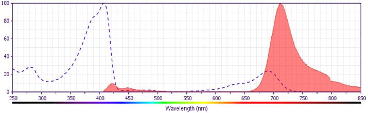
The antibody was conjugated to BD Horizon BV711 which is part of the BD Horizon Brilliant™ Violet family of dyes. This dye is a tandem fluorochrome of BD Horizon BV421 with an Ex Max of 405-nm and an acceptor dye with an Em Max at 711-nm. BD Horizon BV711 can be excited by the violet laser and detected in a filter used to detect Cy™5.5 / Alexa Fluor® 700-like dyes (eg, 712/20-nm filter). Due to the excitation and emission characteristics of the acceptor dye, there may be moderate spillover into the Alexa Fluor® 700 and PerCP-Cy5.5 detectors. However, the spillover can be corrected through compensation as with any other dye combination.
Development References (8)
-
Bruno S, Crissman HA, Bauer KD, Darzynkiewicz Z. Changes in cell nuclei during S phase: progressive chromatin condensation and altered expression of the proliferation-associated nuclear proteins Ki-67, cyclin (PCNA), p105, and p34. Exp Cell Res. 1991; 196(1):99-106. (Biology: Flow cytometry). View Reference
-
Bruno S, Darzynkiewicz Z. Cell cycle dependent expression and stability of the nuclear protein detected by Ki-67 antibody in HL-60 cells. Cell Prolif. 1992; 25(1):31-40. (Biology: Flow cytometry). View Reference
-
Kouro T, Medina KL, Oritani K, Kincade PW. Characteristics of early murine B-lymphocyte precursors and their direct sensitivity to negative regulators. Blood. 2001; 97(9):2708-2715. (Clone-specific: Flow cytometry). View Reference
-
Picker LJ, Hagen SI, Lum R, et al. Insufficient production and tissue delivery of CD4+ memory T cells in rapidly progressive simian immunodeficiency virus infection. J Exp Med. 2004; 200(10):1299-1314. (Clone-specific: Flow cytometry). View Reference
-
Pitcher CJ, Hagen SI, Walker JM, et al. Development and homeostasis of T cell memory in rhesus macaque. J Immunol. 2002; 168(1):29-43. (Clone-specific: Flow cytometry). View Reference
-
Spargo LDJ, Cleland LG, Cockshell MP, Mayrhofer Graham. Recruitment and proliferation of CD4+ T cells in synovium following adoptive transfer of adjuvant-induced arthritis. Int Immunol. 2006; 18(6):897-910. (Clone-specific: Flow cytometry, Immunofluorescence).
-
Starborg M, Gell K, Brundell E, Höög C. The murine Ki-67 cell proliferation antigen accumulates in the nucleolar and heterochromatic regions of interphase cells and at the periphery of the mitotic chromosomes in a process essential for cell cycle progression. J Cell Sci. 1996; 109(1):143-153. (Biology). View Reference
-
Valenti LM, Mathieu J, Chancerelle Y, et al. High levels of endogenous nitric oxide produced after burn injury in rats arrest activated T lymphocytes in the first G1 phase of the cell cycle and then induce their apoptosis. Exp Cell Res. 2005; 306(1):150-167. (Clone-specific: Flow cytometry). View Reference
Please refer to Support Documents for Quality Certificates
Global - Refer to manufacturer's instructions for use and related User Manuals and Technical data sheets before using this products as described
Comparisons, where applicable, are made against older BD Technology, manual methods or are general performance claims. Comparisons are not made against non-BD technologies, unless otherwise noted.
For Research Use Only. Not for use in diagnostic or therapeutic procedures.