
Preparation And Storage
Recommended Assay Procedures
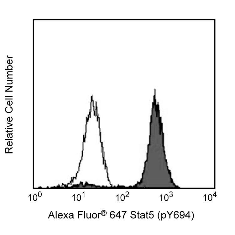
This antibody conjugate is suitable for intracellular staining of human whole blood (using BD Phosflow™ Lyse/Fix Buffer) and peripheral blood mononuclear cells (using BD Cytofix™ Fixation Buffer or BD Phosflow™ Fix Buffer I).
This purified or conjugated mAb was characterized by flow cytometry (Flow) and western blot analysis (WB) using these model systems:
Method Species Cells Treatment Fixation Perm buffer Result
Flow Human PBMC IL-2 Fixation Buffer III Positive Staining
Flow Human PBMC IL-2 Fixation Buffer I or II Unsatisfactory
Flow Human Whole Blood IL-2 Lyse/Fix III Positive Staining
Flow Human Whole Blood IL-2 Lyse/Fix I or II Unsatisfactory
WB Human A431 Cell Lysate EGF Not Applicable Not Applicable 92 kDa
Product Notices
- This reagent has been pre-diluted for use at the recommended Volume per Test. We typically use 1 × 10^6 cells in a 100-µl experimental sample (a test).
- Please refer to www.bdbiosciences.com/us/s/resources for technical protocols.
- Alexa Fluor® 647 fluorochrome emission is collected at the same instrument settings as for allophycocyanin (APC).
- The Alexa Fluor®, Pacific Blue™, and Cascade Blue® dye antibody conjugates in this product are sold under license from Molecular Probes, Inc. for research use only, excluding use in combination with microarrays, or as analyte specific reagents. The Alexa Fluor® dyes (except for Alexa Fluor® 430), Pacific Blue™ dye, and Cascade Blue® dye are covered by pending and issued patents.
- For fluorochrome spectra and suitable instrument settings, please refer to our Multicolor Flow Cytometry web page at www.bdbiosciences.com/colors.
- Source of all serum proteins is from USDA inspected abattoirs located in the United States.
- Caution: Sodium azide yields highly toxic hydrazoic acid under acidic conditions. Dilute azide compounds in running water before discarding to avoid accumulation of potentially explosive deposits in plumbing.
- Alexa Fluor® is a registered trademark of Molecular Probes, Inc., Eugene, OR.
Companion Products


Stat (Signal transducer and activators of transcription) proteins mediate the biological activity of cytokines, including interleukins, interferons, erythropoietin, and growth factors. Ligand-receptor interaction activates constitutively-associated JAK family kinases and subsequent recruitment/activation of Stat proteins by tyrosine phosphorylation. Active Stat proteins then move to the nucleus to promote transcription of cytokine-inducible genes. Seven Stat proteins have been cloned, each of which is differentially expressed and/or activated in a cytokine-specific and cell type-specific manner. Stat5 has been characterized and shown to be encoded by two separate genes, Stat5a and Stat5b, which share over 90% identity at the amino acid level. Stat5a has been shown to be involved in lactogenesis and mammary development, while Stat5b has been shown to be involved in growth hormone signaling and liver gene expression. Both Stat5a and Stat5b are involved in IL-2 induced peripheral T cell proliferation. The peptide hormone, prolactin, binds to the prolactin receptor (PRLR) to initiate the lactogenic response. There are at least three forms of PRLR; however, only the long form activates the 92-kDa Stat5 protein by inducing phosphorylation at Y694. Once phosphorylated, Stat5 becomes an essential transcription factor which binds to the β-casein gene promoter. The presence of an SH2 domain within Stat5 suggests that it may directly interact with protein tyrosine kinases (PTKs) such as JAK2.
The 47 monoclonal antibody recognizes the phosphorylated Y694 of Stat5a. The homologous phosphorylation site in Stat5b is Y699.
Development References (5)
-
Bromberg J, Darnell JE. The role of STATs in transcriptional control and their impact on cellular function. Oncogene. 2000; 19(21):2468-2473. (Biology). View Reference
-
Gouilleux F, Wakao H, Mundt M, Groner B. Prolactin induces phosphorylation of Tyr694 of Stat5 (MGF), a prerequisite for DNA binding and induction of transcription. EMBO J. 1994; 13(18):4361-4369. (Biology). View Reference
-
Imada K, Leonard WJ. The Jak-STAT pathway. Mol Immunol. 2000; 37:1-11. (Biology). View Reference
-
Liu KD, Gaffen SL, Goldsmith MA. JAK/STAT signaling by cytokine receptors. Curr Opin Immunol. 1998; 10(3):271-278. (Biology). View Reference
-
Wakao H, Gouilleux F, Groner B. Mammary gland factor (MGF) is a novel member of the cytokine regulated transcription factor gene family and confers the prolactin response. EMBO J. 1994; 13(9):2182-2191. (Biology). View Reference
Please refer to Support Documents for Quality Certificates
Global - Refer to manufacturer's instructions for use and related User Manuals and Technical data sheets before using this products as described
Comparisons, where applicable, are made against older BD Technology, manual methods or are general performance claims. Comparisons are not made against non-BD technologies, unless otherwise noted.
For Research Use Only. Not for use in diagnostic or therapeutic procedures.