

Preparation And Storage
Recommended Assay Procedures
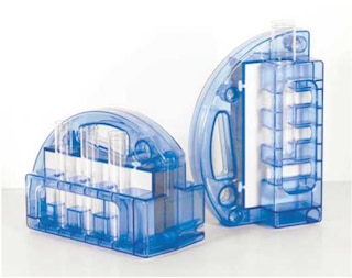
Peripheral Blood Mononuclear Cells (PBMC) are labeled with BD IMag™ Anti-Human CD45RA Particles - DM, then placed within the magnetic field of the BD IMag™ Cell Separation Magnet. Labeled cells migrate toward the magnet (positive fraction), leaving the unlabeled cells in suspension to be drawn off (negative fraction). The tube is then removed from the magnetic field to resuspend the positive fraction and separated twice more to increase the purity of the positive fraction. The magnetic separation steps are diagrammed in the Separation Flow Chart. After the positive fraction is washed, it can be evaluated in downstream applications such as flow cytometry and tissue culture.
MAGNETIC LABELING AND SEPARATION PROTOCOL
1. Dilute BD IMag™ Buffer (10X) (Cat. No. 552362) 1:10 with sterile distilled water or prepare Phosphate Buffered Saline supplemented with 0.5% BSA, 2 mM EDTA, and 0.1% sodium azide), and store at 4°C.
Optional: If only CD45RA-positive T cells are desired, enrich the T lymphocytes by using the BD IMag™ Human T Lymphocyte, CD4 T Lymphocyte, or CD8 T Lymphocyte Enrichment Set - DM (Cat. No. 557874, 557939, or 557941, respectively).
2. Prepare PBMC from anti-coagulated human blood, preferably by density gradient centrifugation using Ficoll-Paque™.*
3. Count the cells, wash them with an excess volume of 1X BD IMag™ buffer, and carefully aspirate all the supernatant.
4. Vortex the BD IMag™ Anti-Human CD45RA Particles - DM thoroughly, and add 50 µl of particles for every 10^7 total cells.
5. MIX THOROUGHLY. Incubate at room temperature for 30 minutes.†
6. Bring the BD IMag™-particle labeling volume up to 1 - 8 x 10^7 cells/ml with 1X BD IMag™ buffer, and immediately place the tube on the Cell Separation Magnet. Incubate for 8 - 10 minutes.
7. With the tube on the Cell Separation Magnet, carefully aspirate off the supernatant. This supernatant contains the negative fraction.
8. Remove the tube from the Cell Separation Magnet, and add 1X BD IMag™ buffer to the same volume as in Step 6. Gently resuspend cells by pipetting up and down, and return the tube to the Cell Separation Magnet for another 2 - 4 minutes.
9. With the tube on the Cell Separation Magnet, carefully aspirate off the supernatant and discard.
10.Repeat Steps 8 and 9.
11.After the final wash step, resuspend the positive fraction in an appropriate buffer or medium, and proceed with desired downstream application(s).
NOTES:
* Hints for successful cell preparation:
o Draw the blood into a tube containing EDTA
o Remove the platelet rich plasma by centrifuging once at 220-240 × g.
o Wash 2-3 times in PBS after the density gradient separation.
o Remove clumps of cells and/or debris by passing the suspension through a 70-µm nylon cell strainer.
† Avoid nonspecific labeling by working quickly and adhering to recommended incubation times.
Product Notices
- Source of all serum proteins is from USDA inspected abattoirs located in the United States.
- Caution: Sodium azide yields highly toxic hydrazoic acid under acidic conditions. Dilute azide compounds in running water before discarding to avoid accumulation of potentially explosive deposits in plumbing.
- For fluorochrome spectra and suitable instrument settings, please refer to our Multicolor Flow Cytometry web page at www.bdbiosciences.com/colors.
- BD IMag™ particles are prepared from carboxy-functionalized magnetic particles which are manufactured by Skold Technology and are licensed under US patent number 7,169,618.
- Please refer to www.bdbiosciences.com/us/s/resources for technical protocols.
Companion Products

BD™ IMag Anti-Human CD45RA Particles - DM are magnetic nanoparticles that have monoclonal antibody conjugated to their surfaces. These particles are optimized for the positive selection or depletion of the CD45RA-bearing leukocytes using the BD IMag™ Cell Separation Magnet. CD45RA is the 220 kDa isoform of the human leukocyte common antigen found on major subsets of peripheral CD4+ and CD8+ T lymphocytes, B lymphocytes, NK cells, and monocytes. CD45RO and CD45RA expression defines complementary, predominantly non-overlapping populations of T cells in peripheral blood; and it is generally accepted that naive T cells are CD45RO- CD45RA+, while memory T cells are CD45RO+ CD45RA-. To specifically enrich CD45RA-expressing naive T lymphocytes, we recommend first depleting the erythrocytes, platelets, and non-T leukocytes, by using the appropriate BD™ IMag human T lymphocyte enrichment set, followed by positive selection of the CD45RA+ population.
Development References (3)
-
Johnson P, Maiti A, Ng DHW. CD45: A family of leukocyte-specific cell surface glycoproteins. In: Herzenberg LA, Weir DM, Herzenberg LA, Blackwell C , ed. Weir's Handbook of Experimental Immunology, Vol 2. Cambridge: Blackwell Science; 1997:62.1-62.16.
-
Picker LJ, Treer JR, Ferguson-Darnell B, Collins PA, Buck D, Terstappen LW. Control of lymphocyte recirculation in man. I. Differential regulation of the peripheral lymph node homing receptor L-selectin on T cells during the virgin to memory cell transition. J Immunol. 1993; 150(3):1105-1121. (Biology). View Reference
-
Schwinzer R. Cluster Report: CD45/CD45R. In: Knapp W. W. Knapp .. et al., ed. Leucocyte typing IV : white cell differentiation antigens. Oxford New York: Oxford University Press; 1989:628-634.
Please refer to Support Documents for Quality Certificates
Global - Refer to manufacturer's instructions for use and related User Manuals and Technical data sheets before using this products as described
Comparisons, where applicable, are made against older BD Technology, manual methods or are general performance claims. Comparisons are not made against non-BD technologies, unless otherwise noted.
For Research Use Only. Not for use in diagnostic or therapeutic procedures.