Skip to main content Skip to navigation
RY586 Rat Anti-Mouse IL-4
Two-color flow cytometric analysis of IL-4 expression in stimulated Mouse T cells. Mouse splenic CD4+ T cells were cultured (2 days) with plate-bound Purified NA/LE Hamster Anti-Mouse CD3e (Cat. No. 553057; 10 μg/ml for coating) and soluble Purified NA/LE Hamster Anti-Mouse CD28 (Cat. No. 553294; 2 μg/ml) antibodies with Recombinant Mouse IL-2 (Cat. No. 550069; 10 ng/ml) and Mouse IL-4 (Cat. No. 550067; 25 ng/ml) proteins. The cells were subsequently cultured (3 days) with Recombinant IL-2 and IL-4. The cells were then stimulated with Phorbol 12-Myristate 13-Acetate (PMA; Sigma Cat. No. P-8139) and Ionomycin (Sigma Cat. No. I-0634) in the presence of BD GolgiStop™ Protein Transport Inhibitor (containing Monensin) (Cat. No. 554724) for 6 hours. The cells were washed and fixed with BD Cytofix™ Fixation Buffer (Cat. No. 554655). The fixed cells were washed and then permeabilized and stained  in BD Perm/Wash™ Buffer (Cat. No. 554723) with APC Rat Anti-Mouse CD4 antibody (Cat. No. 561091/553051) and with either BD Horizon™ RY586 Rat IgG1, κ Isotype Control (Cat. No. 568822; Left Plot) or BD Horizon™ RY586 Rat Anti-Mouse IL-4 antibody (Cat. No. 568541/568542; Right Plot) at 0.125 µg/test using the BD Biosciences Intracellular Cytokine Staining protocol. The bivariate pseudocolor density plot showing the correlated expression of IL-4 (or Ig Isotype control staining) versus CD4 was derived from gated events with the forward and side light-scatter characteristics of intact stimulated lymphocytes. Flow cytometry and data analysis were performed using a BD LSRFortessa™ X-20 Cell Analyzer System and FlowJo™ software. Data shown on this Technical Data Sheet are not lot specific.
Product Details
Down Arrow Up Arrow


BD Horizon™
IL4: Interleukin-4; BSF-1; B-cell growth factor 1; BCGF-1
Mouse (QC Testing)
Rat IgG1, κ
Partially Purified Mouse IL-4
Intracellular staining (flow cytometry) (Routinely Tested)
0.2 mg/ml
16189
Aqueous buffered solution containing ≤0.09% sodium azide.
RUO


Preparation And Storage

Store undiluted at 4°C and protected from prolonged exposure to light. Do not freeze. The monoclonal antibody was purified from tissue culture supernatant or ascites by affinity chromatography. The antibody was conjugated to the dye under optimum conditions and unreacted dye was removed.

Recommended Assay Procedures

BD® CompBeads can be used as surrogates to assess fluorescence spillover (compensation). When fluorochrome conjugated antibodies are bound to BD® CompBeads, they have spectral properties very similar to cells. However, for some fluorochromes there can be small differences in spectral emissions compared to cells, resulting in spillover values that differ when compared to biological controls. It is strongly recommended that when using a reagent for the first time, users compare the spillover on cells and BD® CompBeads to ensure that BD® CompBeads are appropriate for your specific cellular application.

Product Notices

  1. Please refer to www.bdbiosciences.com/us/s/resources for technical protocols.
  2. Caution: Sodium azide yields highly toxic hydrazoic acid under acidic conditions. Dilute azide compounds in running water before discarding to avoid accumulation of potentially explosive deposits in plumbing.
  3. Since applications vary, each investigator should titrate the reagent to obtain optimal results.
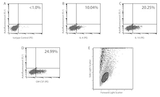
  4. An isotype control should be used at the same concentration as the antibody of interest.
  5. For fluorochrome spectra and suitable instrument settings, please refer to our Multicolor Flow Cytometry web page at www.bdbiosciences.com/colors.
  6. Please refer to http://regdocs.bd.com to access safety data sheets (SDS).
  7. CF™ is a trademark of Biotium, Inc.
568541 Rev. 1
Antibody Details
Down Arrow Up Arrow
11B11

Interleukin-4 (IL-4) is a pleiotropic cytokine that has many roles, such as inducing the differentiation of naïve helper T cells (Th0 cells) to Th2 cells, stimulating activated B-cell and T-cell proliferation, and promoting immunoglobulin class switching to IgG1 and IgE in mouse B-cells.  IL-4 is expressed by CD4 T-cells, mast cells, basophils and eosinophils.  IL-4 was previously known as B-Cell Differentiation Factor (BCDF) or B-cell Stimulatory Factor (BSF1).  The 11B11 monoclonal antibody specifically binds to mouse IL-4. The immunogen used to generate the 11B11 hybridoma was partially purified mouse IL-4 prepared from the supernatant of Phorbol 12-Myristate 13-Acetate (PMA)-stimulated EL-4 cells. The 11B11 antibody is reportedly a neutralizing antibody.

568541 Rev. 1
Format Details
Down Arrow Up Arrow
RY586
The BD Horizon RealYellow™ 586 (RY586) Dye is part of the BD family of yellow-green dyes. It is a small organic fluorochrome with an excitation maximum (Ex Max) at 565-nm and an emission maximum (Em Max) at 586-nm. Driven by BD innovation, RY586 can be used on both spectral and conventional cytometers and is designed to be excited by the Yellow-Green laser (561-nm) with minimal excitation by the 488-nm Blue laser. For conventional instruments equipped with a Yellow-Green laser (561-nm), RY586 can be used as an alternative to PE and we recommend using an optical filter centered near 586-nm (eg, a 586/15-nm bandpass filter). For spectral instruments equipped with a Yellow-Green laser (561-nm), it can be used in conjunction with PE. Compared to PE, RY586 is similar in brightness, minimal spillover into Blue detectors, and increased spillover into the 610/20-nm (PE-CF594) detector. Please ensure that your instrument configuration (lasers and optical filters) is appropriate for this dye.
altImg
RY586
Yellow-Green 561 nm
564 nm
586 nm
568541 Rev.1
Citations & References
Down Arrow Up Arrow

Development References (10)

  1. Abrams J. Immunoenzymetric assay of mouse and human cytokines using NIP-labeled anti-cytokine antibodies. Curr Protoc Immunol. 2001; 1:6.20-6.21. (Clone-specific: ELISA). View Reference
  2. Assenmacher M, Schmitz J, Radbruch A. Flow cytometric determination of cytokines in activated murine T helper lymphocytes: expression of interleukin-10 in interferon-gamma and in interleukin-4-expressing cells. Eur J Immunol. 1994; 24(5):1097-1101. (Clone-specific: Flow cytometry). View Reference
  3. Haak-Frendscho M, Brown JF, Iizawa Y, Wagner RD, Czuprynski CJ. Administration of anti-IL-4 monoclonal antibody 11B11 increases the resistance of mice to Listeria monocytogenes infection. J Immunol. 1992; 148(12):3978-3985. (Clone-specific: Neutralization). View Reference
  4. Lindqvist C, Lundstrom H, Oker-Blom C, Akerman KE. Enhanced IL-4-mediated D10.G4.1 proliferation with suboptimal concentrations of anti-IL-4 receptor monoclonal antibodies. J Immunol. 1993; 150(2):394-398. (Clone-specific: Neutralization). View Reference
  5. Ohara J, Paul WE. Production of a monoclonal antibody to and molecular characterization of B-cell stimulatory factor-1. Nature. 1985; 315(6017):333-336. (Immunogen). View Reference
  6. Openshaw P, Murphy EE, Hosken NA, et al. Heterogeneity of intracellular cytokine synthesis at the single-cell level in polarized T helper 1 and T helper 2 populations. J Exp Med. 1995; 182(5):1357-1367. (Clone-specific: Flow cytometry). View Reference
  7. Prussin C, Metcalfe DD. Detection of intracytoplasmic cytokine using flow cytometry and directly conjugated anti-cytokine antibodies. J Immunol Methods. 1995; 188(1):117-128. (Methodology). View Reference
  8. Sadick MD, Heinzel FP, Holaday BJ, Pu RT, Dawkins RS, Locksley RM. Cure of murine leishmaniasis with anti-interleukin 4 monoclonal antibody. Evidence for a T cell-dependent, interferon gamma-independent mechanism. J Exp Med. 1990; 171(1):115-127. (Clone-specific: Neutralization). View Reference
  9. Sander B, Hoiden I, Andersson U, Moller E, Abrams JS. Similar frequencies and kinetics of cytokine producing cells in murine peripheral blood and spleen. Cytokine detection by immunoassay and intracellular immunostaining. J Immunol Methods. 1993; 166(2):201-214. (Clone-specific: ELISA, Flow cytometry). View Reference
  10. Swain SL, Weinberg AD, English M, Huston G. IL-4 directs the development of Th2-like helper effectors. J Immunol. 1990; 145(11):3796-3806. (Clone-specific: Neutralization). View Reference
View All (10) View Less
568541 Rev. 1

Please refer to Support Documents for Quality Certificates


Global - Refer to manufacturer's instructions for use and related User Manuals and Technical data sheets before using this products as described


Comparisons, where applicable, are made against older BD Technology, manual methods or are general performance claims.  Comparisons are not made against non-BD technologies, unless otherwise noted.

For Research Use Only. Not for use in diagnostic or therapeutic procedures.