Preparation And Storage
Recommended Assay Procedures
BD® CompBeads can be used as surrogates to assess fluorescence spillover (compensation). When fluorochrome conjugated antibodies are bound to BD® CompBeads, they have spectral properties very similar to cells. However, for some fluorochromes there can be small differences in spectral emissions compared to cells, resulting in spillover values that differ when compared to biological controls. It is strongly recommended that when using a reagent for the first time, users compare the spillover on cells and BD® CompBeads to ensure that BD® CompBeads are appropriate for your specific cellular application.
Product Notices
- Researchers should determine the optimal concentration of this reagent for their individual applications.
- The production process underwent stringent testing and validation to assure that it generates a high-quality conjugate with consistent performance and specific binding activity. However, verification testing has not been performed on all conjugate lots.
- Please refer to www.bdbiosciences.com/us/s/resources for technical protocols.
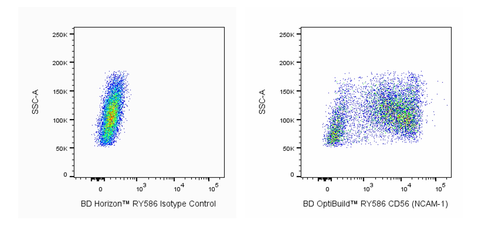
- An isotype control should be used at the same concentration as the antibody of interest.
- Caution: Sodium azide yields highly toxic hydrazoic acid under acidic conditions. Dilute azide compounds in running water before discarding to avoid accumulation of potentially explosive deposits in plumbing.
- CF™ is a trademark of Biotium, Inc.
- Please refer to http://regdocs.bd.com to access safety data sheets (SDS).
- For fluorochrome spectra and suitable instrument settings, please refer to our Multicolor Flow Cytometry web page at www.bdbiosciences.com/colors.
- Since applications vary, each investigator should titrate the reagent to obtain optimal results.
Companion Products


The 809220 monoclonal antibody specifically recognizes CD56 which is also known as Neural cell adhesion molecule 1 (NCAM-1), Neural cell adhesion molecule (NCAM), or embryonic NCAM (E-NCAM). CD56 (NCAM-1) is a 120-180 kDa type I transmembrane glycoprotein that is encoded by Ncam1 which belongs to the immunoglobulin superfamily (IgSF). Three different isoforms of CD56 (NCAM-1) have been described that vary in their cytoplasmic domains. The 180 kDa long isoform contains five consecutive IgC-like domains followed by two fibronectin type III domains in its extracellular region, a transmembrane sequence and a large cytoplasmic domain. A 140 kDa isoform has a shorter cytoplasmic tail whereas the 120 kDa isoform is glycophosphatidylinositol (GPI)-linked to the cell membrane. CD56 (NCAM-1) functions on cells as a hemophilic or heterophilic adhesion molecule and as a receptor for various ligands including certain growth factors. Throughout development and adulthood, CD56 (NCAM-1) is expressed on various cell types such as neurons where it might play roles in cellular migration, axonal guidance, and synapse formation. It can also be expressed on astrocytes as well as bone marrow neutrophils and some monocytes and might be involved in cellular migration and other functions. Polysialylation of CD56 (NCAM-1) reduces its ability to dimerize and can thereby affect the functions of cells that express it.

Development References (4)
-
Fujita T, Chen MJ, Li B, et al. Neuronal transgene expression in dominant-negative SNARE mice.. J Neurosci. 2014; 34(50):16594-604. (Clone-specific: Fluorescence activated cell sorting). View Reference
-
Li S, Nie EH, Yin Y, et al. GDF10 is a signal for axonal sprouting and functional recovery after stroke.. Nat Neurosci. 2015; 18(12):1737-45. (Clone-specific: Fluorescence activated cell sorting). View Reference
-
Rougon G, Deagostini-Bazin H, Hirn M, Goridis C. Tissue- and developmental stage-specific forms of a neural cell surface antigen linked to differences in glycosylation of a common polypeptide.. EMBO J. 1982; 1(10):1239-44. (Biology). View Reference
-
Stamatos NM, Zhang L, Jokilammi A, et al. Changes in polysialic acid expression on myeloid cells during differentiation and recruitment to sites of inflammation: role in phagocytosis. Glycobiology. 2014; 24(9):864-879. (Biology). View Reference
Please refer to Support Documents for Quality Certificates
Global - Refer to manufacturer's instructions for use and related User Manuals and Technical data sheets before using this products as described
Comparisons, where applicable, are made against older BD Technology, manual methods or are general performance claims. Comparisons are not made against non-BD technologies, unless otherwise noted.
For Research Use Only. Not for use in diagnostic or therapeutic procedures.