Preparation And Storage
Recommended Assay Procedures
BD® CompBeads can be used as surrogates to assess fluorescence spillover (compensation). When fluorochrome conjugated antibodies are bound to BD® CompBeads, they have spectral properties very similar to cells. However, for some fluorochromes there can be small differences in spectral emissions compared to cells, resulting in spillover values that differ when compared to biological controls. It is strongly recommended that when using a reagent for the first time, users compare the spillover on cells and BD® CompBeads to ensure that BD® CompBeads are appropriate for your specific cellular application.
Product Notices
- Please refer to www.bdbiosciences.com/us/s/resources for technical protocols.
- Caution: Sodium azide yields highly toxic hydrazoic acid under acidic conditions. Dilute azide compounds in running water before discarding to avoid accumulation of potentially explosive deposits in plumbing.
- Since applications vary, each investigator should titrate the reagent to obtain optimal results.
- For fluorochrome spectra and suitable instrument settings, please refer to our Multicolor Flow Cytometry web page at www.bdbiosciences.com/colors.
- An isotype control should be used at the same concentration as the antibody of interest.
- Human donor specific background has been observed in relation to the presence of anti-polyethylene glycol (PEG) antibodies, developed as a result of certain vaccines containing PEG, including some COVID-19 vaccines. We recommend use of BD Horizon Brilliant™ Stain Buffer in your experiments to help mitigate potential background. For more information visit https://www.bdbiosciences.com/en-us/support/product-notices.
- Species cross-reactivity detected in product development may not have been confirmed on every format and/or application.
- CF™ is a trademark of Biotium, Inc.
- Please refer to http://regdocs.bd.com to access safety data sheets (SDS).
Companion Products


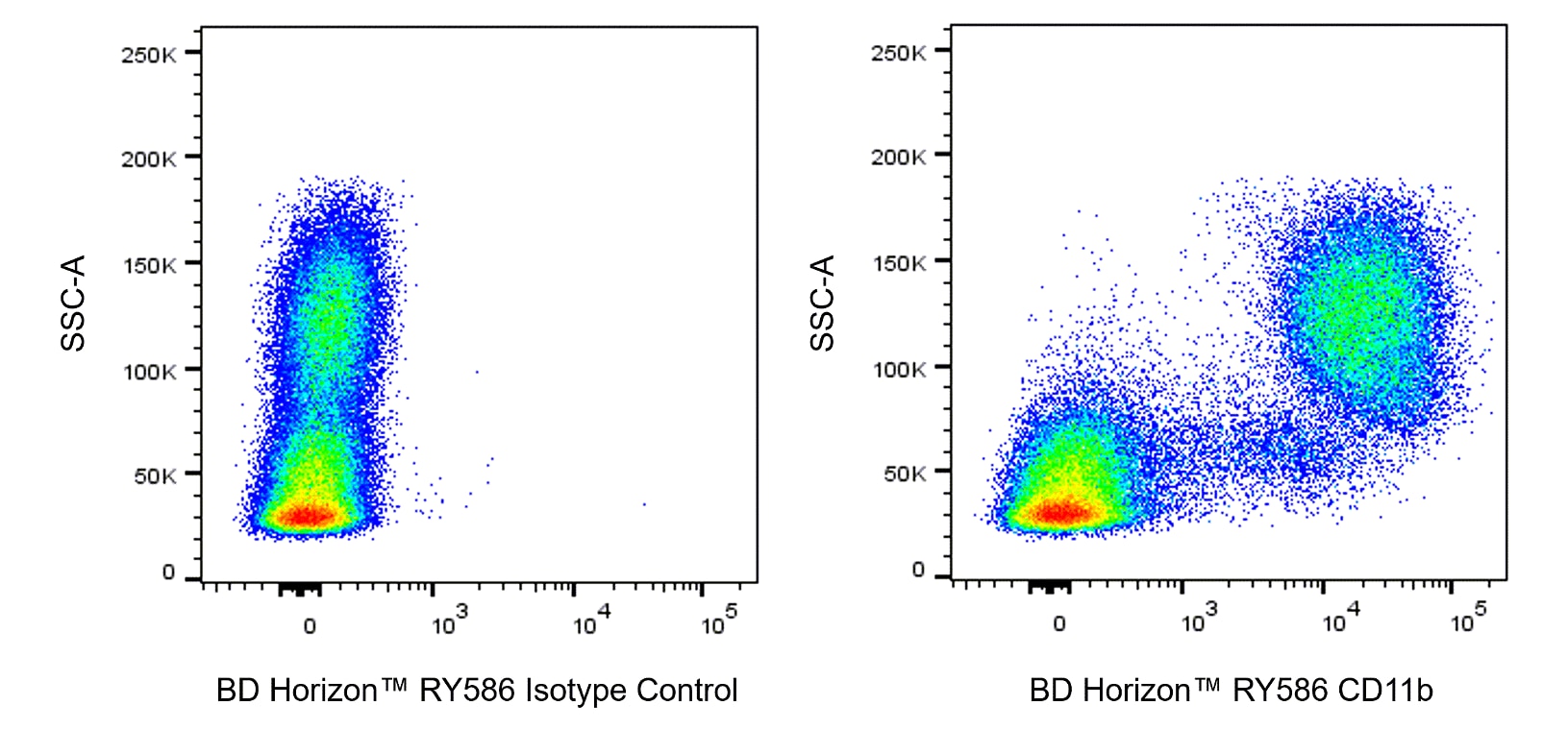
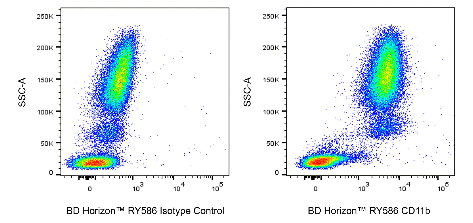
The M1/70 monoclonal antibody specifically binds to CD11b, also known as Integrin alpha M (Itgam or αM). CD11b is a 170-kDa type 1 transmembrane glycoprotein and belongs to the Integrin alpha chain family. CD11b serves as the alpha chain of the heterodimeric Mac-1 integrin (CD11b/CD18, αMβ2), also known as complement receptor 3 (CR3). Mac-1 mediates adhesion to ICAM-1 (CD54), ICAM-2 (CD102), fibrinogen and binding to C3bi. Mac-1 is expressed at varying levels on granulocytes, macrophages, myeloid-derived dendritic cells, natural killer cells, microglia, and B-1 B lymphocytes. Mac-1 expression is rapidly upregulated on neutrophils after activation, in the same time period that CD62L (L-selectin) is shed from the cell surface. The M1/70 antibody reportedly blocks cell adherence and C3bi binding but does not block cell-mediated lysis. Cross-reaction of the M1/70 antibody with CD11b expressed on human monocytes, polymorphonuclear leukocytes, and NK cells has been reported.

Development References (11)
-
Ault KA, Springer TA. Cross-reaction of a rat-anti-mouse phagocyte-specific monoclonal antibody (anti-Mac-1) with human monocytes and natural killer cells. J Immunol. 1981; 126(1):359-364. (Clone-specific: Flow cytometry, Fluorescence activated cell sorting, Radioimmunoassay). View Reference
-
Beller DI, Springer TA, Schreiber RD. Anti-Mac-1 selectively inhibits the mouse and human type three complement receptor. J Exp Med. 1982; 156(4):1000-1009. (Clone-specific: Blocking, Inhibition). View Reference
-
Driver DJ, McHeyzer-Williams LJ, Cool M, Stetson DB, McHeyzer-Williams MG. Development and maintenance of a B220- memory B cell compartment. J Immunol. 2001; 167(3):1393-1405. (Clone-specific: Flow cytometry, Immunofluorescence). View Reference
-
Kaji K, Takeshita S, Miyake K, Takai T, Kudo A. Functional association of CD9 with the Fc gamma receptors in macrophages. J Immunol. 2001; 166(5):3256-3265. (Clone-specific: Fluorescence microscopy, Immunofluorescence). View Reference
-
Lagasse E, Weissman IL. Flow cytometric identification of murine neutrophils and monocytes. J Immunol Methods. 1996; 197(1-2):139-150. (Clone-specific: Flow cytometry). View Reference
-
Lub M, van Kooyk Y, Figdor CG. Competition between lymphocyte function-associated antigen 1 (CD11a/CD18) and Mac-1 (CD11b/CD18) for binding to intercellular adhesion molecule-1 (CD54). J Leukoc Biol. 1996; 59(5):648-655. (Clone-specific: Immunoprecipitation). View Reference
-
Sanchez-Madrid F, Simon P, Thompson S, Springer TA. Mapping of antigenic and functional epitopes on the alpha- and beta-subunits of two related mouse glycoproteins involved in cell interactions, LFA-1 and Mac-1. J Exp Med. 1983; 158(2):586-602. (Clone-specific: Immunoaffinity chromatography, Immunoprecipitation, Inhibition). View Reference
-
Springer T, Galfre G, Secher D, Milstein C. Monoclonal xenogeneic antibodies to mouse leukocyte antigens: identification of macrophage-specific and other differentiation antigens. Curr Top Microbiol Immunol. 1978; 81:45-50. (Immunogen: Immunoprecipitation, Radioimmunoassay). View Reference
-
Springer T, Galfre G, Secher DS, Milstein C. Mac-1: a macrophage differentiation antigen identified by monoclonal antibody. Eur J Immunol. 1979; 9(4):301-306. (Clone-specific: Flow cytometry, Immunoprecipitation). View Reference
-
Springer T, Galfre G, Secher DS, Milstein C. Monoclonal xenogeneic antibodies to murine cell surface antigens: identification of novel leukocyte differentiation antigens. Eur J Immunol. 1978; 8(8):539-551. (Immunogen: Immunoprecipitation). View Reference
-
Springer TA, Davignon D, Ho MK, Kurzinger K, Martz E, Sanchez-Madrid F. LFA-1 and Lyt-2,3, molecules associated with T lymphocyte-mediated killing; and Mac-1, an LFA-1 homologue associated with complement receptor function. Immunol Rev. 1982; 68:171-195. (Immunogen: Immunoprecipitation, Radioimmunoassay). View Reference
Please refer to Support Documents for Quality Certificates
Global - Refer to manufacturer's instructions for use and related User Manuals and Technical data sheets before using this products as described
Comparisons, where applicable, are made against older BD Technology, manual methods or are general performance claims. Comparisons are not made against non-BD technologies, unless otherwise noted.
For Research Use Only. Not for use in diagnostic or therapeutic procedures.