Preparation And Storage
Recommended Assay Procedures
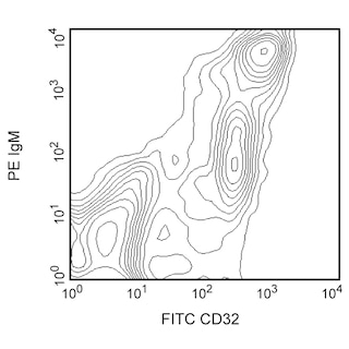
BD® CompBeads can be used as surrogates to assess fluorescence spillover (compensation). When fluorochrome conjugated antibodies are bound to BD® CompBeads, they have spectral properties very similar to cells. However, for some fluorochromes there can be small differences in spectral emissions compared to cells, resulting in spillover values that differ when compared to biological controls. It is strongly recommended that when using a reagent for the first time, users compare the spillover on cells and BD® CompBeads to ensure that BD® CompBeads are appropriate for your specific cellular application.
Product Notices
- Researchers should determine the optimal concentration of this reagent for their individual applications.
- The production process underwent stringent testing and validation to assure that it generates a high-quality conjugate with consistent performance and specific binding activity. However, verification testing has not been performed on all conjugate lots.
- Please refer to www.bdbiosciences.com/us/s/resources for technical protocols.
- An isotype control should be used at the same concentration as the antibody of interest.
- Caution: Sodium azide yields highly toxic hydrazoic acid under acidic conditions. Dilute azide compounds in running water before discarding to avoid accumulation of potentially explosive deposits in plumbing.
- CF™ is a trademark of Biotium, Inc.
- Please refer to http://regdocs.bd.com to access safety data sheets (SDS).
- For fluorochrome spectra and suitable instrument settings, please refer to our Multicolor Flow Cytometry web page at www.bdbiosciences.com/colors.
- Since applications vary, each investigator should titrate the reagent to obtain optimal results.
Companion Products


The OX-49 antibody reacts with the glycoprotein CD44H (also known as CD44s) expressed on most leukocytes, except for a subset of B lymphocytes, and at greatly increased levels on T- and B-cell blasts. The epitope recognized by OX-49 antibody has been mapped to a region on both the standard, CD44s, and the splice variant, CD44v, isoforms of CD44. However, recent reports indicate that OX-49 antibody cannot detect the CD44v isoform, possibly due to conformational changes in the epitope. CD44 is a cell adhesion receptor, and its ligand, hyaluronate, is a common component of extracellular matrices.

Development References (7)
-
Arch R, Wirth K, Hofmann M, et al. Participation in normal immune responses of a metastasis-inducing splice variant of CD44. Science. 1992; 257(5070):682-685. (Biology). View Reference
-
Mitnacht R, Tacke M, Hunig T. Expression of cell interaction molecules by immature rat thymocytes during passage through the CD4+8+ compartment: developmental regulation and induction by T cell receptor engagement of CD2, CD5, CD28, CD11a, CD44 and CD53. Eur J Immunol. 1995; 25(2):328-332. (Biology). View Reference
-
Noonan KJ, Stevens JW, Tammi R, Tammi M, Hernandez JA, Midura RJ. Spatial distribution of CD44 and hyaluronan in the proximal tibia of the growing rat. J Orthop Res. 1996; 14(4):573-581. (Biology). View Reference
-
Paterson DJ, Jefferies WA, Green JR. Antigens of activated rat T lymphocytes including a molecule of 50,000 Mr detected only on CD4 positive T blasts. Mol Immunol. 1987; 24(12):1281-1290. (Immunogen). View Reference
-
Stevens JW, Noonan KJ, Bosch PP, et al. CD44 in growing normal and neoplastic rat cartilage. Ann N Y Acad Sci. 1996; 785:333-336. (Biology). View Reference
-
Westermann J, Nagahori Y, Walter S, Heerwagen C, Miyasaka M, Pabst R. B and T lymphocyte subsets enter peripheral lymph nodes and Peyer's patches without preference in vivo: no correlation occurs between their localization in different types of high endothelial venules and the expression of CD44, VLA-4, LFA-1, ICAM-1, CD2 or L-selectin. Eur J Immunol. 1994; 24(10):2312-2316. (Biology). View Reference
-
Zheng Z, Katoh S, He Q, et al. Monoclonal antibodies to CD44 and their influence on hyaluronan recognition. J Cell Biol. 1995; 130(2):485-495. (Biology). View Reference
Please refer to Support Documents for Quality Certificates
Global - Refer to manufacturer's instructions for use and related User Manuals and Technical data sheets before using this products as described
Comparisons, where applicable, are made against older BD Technology, manual methods or are general performance claims. Comparisons are not made against non-BD technologies, unless otherwise noted.
For Research Use Only. Not for use in diagnostic or therapeutic procedures.