Skip to main content Skip to navigation
RY586 Mouse Anti-Human TCRαβ
Multicolor flow cytometric analysis of TCRαβ expression on Human peripheral blood lymphocytes. Human whole blood was stained with BD Horizon™ BUV395 Mouse Anti-Human CD19 antibody (Cat. No. 563549/563551), BD Horizon™ BV421 Mouse Anti-Human CD3 antibody (Cat. No. 562426/562427) and either BD Horizon™ RY586 Mouse IgG1, κ Isotype Control (Cat No. 568097; Left Plots) or BD Horizon™ RY586 Mouse Anti-Human TCRαβ antibody (Cat. No. 568516/568517; Right Plots). Erythrocytes were lysed with BD FACS Lysing™ Solution (Cat. No. 349202). Flow cytometry and data analysis were performed using a LSRFortessa™ X-20 Flow Cytometer System and FlowJo™ software.              Upper Plots: Bivariate pseudocolor density plots showing the correlated expression of TCRαβ (or Ig Isotype control staining) versus CD19 were derived from gated events with the forward and side light-scatter characteristics of intact lymphocytes.      Bottom Plots: Bivariate pseudocolor density plots showing the correlated expression of TCRαβ (or Ig Isotype control staining) versus CD3 were derived from gated events with the forward and side light-scatter characteristics of intact lymphocytes.
Product Details
Down Arrow Up Arrow


BD Horizon™
alpha-beta T cell receptor, TCRAB, TRA@/TRB@; IP26A
Human (QC Testing)
Mouse BALB/c IgG1, κ
Human T Cell Clone
Flow cytometry (Routinely Tested)
5 µl
Aqueous buffered solution containing ≤0.09% sodium azide.
RUO


Preparation And Storage

Store undiluted at 4°C and protected from prolonged exposure to light. Do not freeze. The monoclonal antibody was purified from tissue culture supernatant or ascites by affinity chromatography. The antibody was conjugated to the dye under optimum conditions and unreacted dye was removed.

Recommended Assay Procedures

BD® CompBeads can be used as surrogates to assess fluorescence spillover (compensation). When fluorochrome conjugated antibodies are bound to BD® CompBeads, they have spectral properties very similar to cells. However, for some fluorochromes there can be small differences in spectral emissions compared to cells, resulting in spillover values that differ when compared to biological controls. It is strongly recommended that when using a reagent for the first time, users compare the spillover on cells and BD® CompBeads to ensure that BD® CompBeads are appropriate for your specific cellular application.

Product Notices

  1. Please refer to www.bdbiosciences.com/us/s/resources for technical protocols.
  2. Caution: Sodium azide yields highly toxic hydrazoic acid under acidic conditions. Dilute azide compounds in running water before discarding to avoid accumulation of potentially explosive deposits in plumbing.
  3. This reagent has been pre-diluted for use at the recommended Volume per Test. We typically use 1 × 10^6 cells in a 100-µl experimental sample (a test).
  4. An isotype control should be used at the same concentration as the antibody of interest.
  5. For fluorochrome spectra and suitable instrument settings, please refer to our Multicolor Flow Cytometry web page at www.bdbiosciences.com/colors.
  6. Human donor specific background has been observed in relation to the presence of anti-polyethylene glycol (PEG) antibodies, developed as a result of certain vaccines containing PEG, including some COVID-19 vaccines. We recommend use of BD Horizon Brilliant™ Stain Buffer in your experiments to help mitigate potential background. For more information visit https://www.bdbiosciences.com/en-us/support/product-notices.
  7. Please refer to http://regdocs.bd.com to access safety data sheets (SDS).
  8. CF™ is a trademark of Biotium, Inc.
568516 Rev. 1
Antibody Details
Down Arrow Up Arrow
IP26

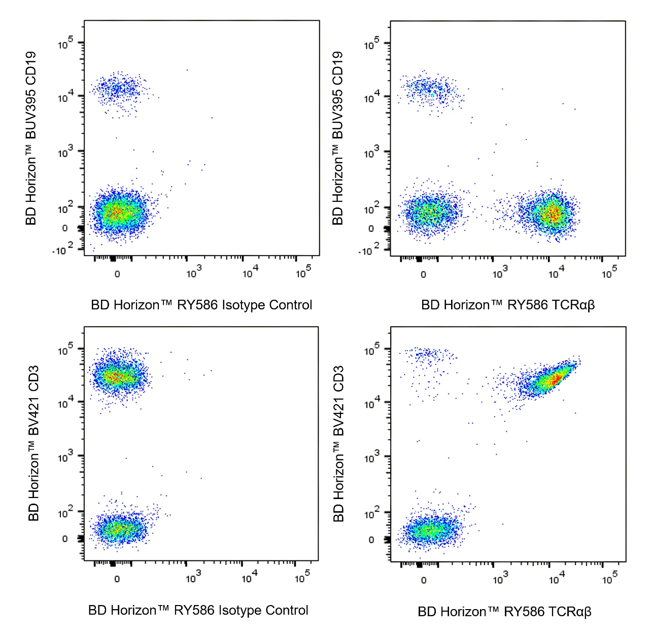
The IP26 monoclonal antibody specifically recognizes a monomorphic determinant on the αβ T-cell receptor (TCRαβ). The TCRαβ is a disulfide-linked 80 kDa heterodimer consisting of a 44 kDa α chain and a 37 kDa β chain. The TCRαβ is normally expressed on 50-70% of thymocytes and on a large fraction of mature T cells including greater than 95% of peripheral blood CD3+ T cells. The TCRαβ serves as a receptor for peptide antigens bound to MHC molecules. The IP26 antibody is mitogenic and useful for the flow cytometric analysis of TCRαβ+ T-cell subsets.

568516 Rev. 1
Format Details
Down Arrow Up Arrow
RY586
The BD Horizon RealYellow™ 586 (RY586) Dye is part of the BD family of yellow-green dyes. It is a small organic fluorochrome with an excitation maximum (Ex Max) at 565-nm and an emission maximum (Em Max) at 586-nm. Driven by BD innovation, RY586 can be used on both spectral and conventional cytometers and is designed to be excited by the Yellow-Green laser (561-nm) with minimal excitation by the 488-nm Blue laser. For conventional instruments equipped with a Yellow-Green laser (561-nm), RY586 can be used as an alternative to PE and we recommend using an optical filter centered near 586-nm (eg, a 586/15-nm bandpass filter). For spectral instruments equipped with a Yellow-Green laser (561-nm), it can be used in conjunction with PE. Compared to PE, RY586 is similar in brightness, minimal spillover into Blue detectors, and increased spillover into the 610/20-nm (PE-CF594) detector. Please ensure that your instrument configuration (lasers and optical filters) is appropriate for this dye.
altImg
RY586
Yellow-Green 561 nm
564 nm
586 nm
568516 Rev.1
Citations & References
Down Arrow Up Arrow

Development References (5)

  1. Agrawal SG, Marquet J, Plumas J, et al. Multiple co-stimulatory signals are required for triggering proliferation of T cells from human secondary lymphoid tissue. Int Immunol. 2001; 13(4):441-450. (Clone-specific: Activation, Functional assay, Stimulation). View Reference
  2. Nikolova M, Marie-Cardine A, Boumsell L, Bensussan A. BY55/CD160 acts as a co-receptor in TCR signal transduction of a human circulating cytotoxic effector T lymphocyte subset lacking CD28 expression. Int Immunol. 2002; 14(5):445-451. (Clone-specific: Flow cytometry). View Reference
  3. Oettgen HC, Kappler J, Tax WJ, Terhorst C. Characterization of the two heavy chains of the T3 complex on the surface of human T lymphocytes. J Biol Chem. 1984; 259(19):12039-12048. (Biology). View Reference
  4. Ortonne N, Huet D, Gaudez C, et al. Significance of circulating T-cell clones in Sezary syndrome. Blood. 2006; 107(10):4030-4038. (Clone-specific: Flow cytometry, Fluorescence activated cell sorting). View Reference
  5. van Dongen JJ, Lhermitte L, Böttcher S, et al. EuroFlow antibody panels for standardized n-dimensional flow cytometric immunophenotyping of normal, reactive and malignant leukocytes. Leukemia. 2012; 26(9):1908-1975. (Clone-specific: Flow cytometry). View Reference
View All (5) View Less
568516 Rev. 1

Please refer to Support Documents for Quality Certificates


Global - Refer to manufacturer's instructions for use and related User Manuals and Technical data sheets before using this products as described


Comparisons, where applicable, are made against older BD Technology, manual methods or are general performance claims.  Comparisons are not made against non-BD technologies, unless otherwise noted.

For Research Use Only. Not for use in diagnostic or therapeutic procedures.