Skip to main content Skip to navigation
RY586 Mouse Anti-Human CD90
Multicolor flow cytometric analysis of CD90 expression on Human peripheral blood lymphocytes. Human peripheral blood mononuclear cells (PBMC) were stained with APC Mouse Anti-Human CD34 antibody (Cat. No. 555824) and with either BD Horizon™ RY586 Mouse IgG1, κ Isotype Control (Cat. No. 568097; Left Plot) or BD Horizon™ RY586 Mouse Anti-Human CD90 antibody (Cat. No. 568495/568496; Right Plot). DAPI (4',6-Diamidino-2-Phenylindole, Dihydrochloride) Solution (Cat. No. 564907) was added to cells right before analysis. The bivariate pseudocolor density plot showing the correlated expression of CD90 (or Ig Isotype control staining) versus CD34 was derived from gated events with the forward and side light-scatter characteristics of viable (DAPI-negative) lymphocytes. Flow cytometry and data analysis were performed using a BD LSRFortessa™ Cell Analyzer System and FlowJo™ software.
RY586 Mouse Anti-Human CD90
Flow cytometric analysis of CD90 expression on Human HEL cells. HEL cells (Human erythroleukemia cell line; ATCC® TIB-180™) were stained with BD Horizon™ RY586 Mouse IgG1, κ Isotype Control (Cat. No. 568097; dashed line histogram) or BD Horizon™ RY586 Mouse Anti-Human CD90 antibody (Cat. No. 568495/568496, solid line histogram). DAPI (4',6-Diamidino-2-Phenylindole, Dihydrochloride) Solution (Cat. No. 564907) was added to cells right before analysis. The fluorescence histogram showing CD90 expression (or Ig Isotype control staining) was derived from gated events with the forward and side light-scatter characteristics of viable (DAPI-negative) cells. Flow cytometry and data analysis were performed using a BD LSRFortessa™ Cell Analyzer System and FlowJo™ software.
Multicolor flow cytometric analysis of CD90 expression on Human peripheral blood lymphocytes. Human peripheral blood mononuclear cells (PBMC) were stained with APC Mouse Anti-Human CD34 antibody (Cat. No. 555824) and with either BD Horizon™ RY586 Mouse IgG1, κ Isotype Control (Cat. No. 568097; Left Plot) or BD Horizon™ RY586 Mouse Anti-Human CD90 antibody (Cat. No. 568495/568496; Right Plot). DAPI (4',6-Diamidino-2-Phenylindole, Dihydrochloride) Solution (Cat. No. 564907) was added to cells right before analysis. The bivariate pseudocolor density plot showing the correlated expression of CD90 (or Ig Isotype control staining) versus CD34 was derived from gated events with the forward and side light-scatter characteristics of viable (DAPI-negative) lymphocytes. Flow cytometry and data analysis were performed using a BD LSRFortessa™ Cell Analyzer System and FlowJo™ software.
Flow cytometric analysis of CD90 expression on Human HEL cells. HEL cells (Human erythroleukemia cell line; ATCC® TIB-180™) were stained with BD Horizon™ RY586 Mouse IgG1, κ Isotype Control (Cat. No. 568097; dashed line histogram) or BD Horizon™ RY586 Mouse Anti-Human CD90 antibody (Cat. No. 568495/568496, solid line histogram). DAPI (4',6-Diamidino-2-Phenylindole, Dihydrochloride) Solution (Cat. No. 564907) was added to cells right before analysis. The fluorescence histogram showing CD90 expression (or Ig Isotype control staining) was derived from gated events with the forward and side light-scatter characteristics of viable (DAPI-negative) cells. Flow cytometry and data analysis were performed using a BD LSRFortessa™ Cell Analyzer System and FlowJo™ software.
Product Details
Down Arrow Up Arrow


BD Horizon™
THY1; Thy-1 antigen; Thy-1 membrane glycoprotein
Human (QC Testing), Rhesus,Cynomolgus,Baboon (Tested in Development)
Mouse BALB/c IgG1, κ
Human HEL Cell Line
Flow cytometry (Routinely Tested)
5 µl
V M07, BP222; VI BP28, E046
7070
Aqueous buffered solution containing ≤0.09% sodium azide.
RUO


Preparation And Storage

Store undiluted at 4°C and protected from prolonged exposure to light. Do not freeze. The monoclonal antibody was purified from tissue culture supernatant or ascites by affinity chromatography. The antibody was conjugated to the dye under optimum conditions and unreacted dye was removed.

Recommended Assay Procedures

BD® CompBeads can be used as surrogates to assess fluorescence spillover (compensation). When fluorochrome conjugated antibodies are bound to BD® CompBeads, they have spectral properties very similar to cells. However, for some fluorochromes there can be small differences in spectral emissions compared to cells, resulting in spillover values that differ when compared to biological controls. It is strongly recommended that when using a reagent for the first time, users compare the spillover on cells and BD® CompBeads to ensure that BD® CompBeads are appropriate for your specific cellular application.

Product Notices

  1. Please refer to www.bdbiosciences.com/us/s/resources for technical protocols.
  2. This reagent has been pre-diluted for use at the recommended Volume per Test. We typically use 1 × 10^6 cells in a 100-µl experimental sample (a test).
  3. An isotype control should be used at the same concentration as the antibody of interest.
  4. Caution: Sodium azide yields highly toxic hydrazoic acid under acidic conditions. Dilute azide compounds in running water before discarding to avoid accumulation of potentially explosive deposits in plumbing.
  5. For fluorochrome spectra and suitable instrument settings, please refer to our Multicolor Flow Cytometry web page at www.bdbiosciences.com/colors.
  6. Human donor specific background has been observed in relation to the presence of anti-polyethylene glycol (PEG) antibodies, developed as a result of certain vaccines containing PEG, including some COVID-19 vaccines. We recommend use of BD Horizon Brilliant™ Stain Buffer in your experiments to help mitigate potential background. For more information visit https://www.bdbiosciences.com/en-us/support/product-notices.
  7. Species cross-reactivity detected in product development may not have been confirmed on every format and/or application.
  8. Please refer to http://regdocs.bd.com to access safety data sheets (SDS).
  9. CF™ is a trademark of Biotium, Inc.
568496 Rev. 2
Antibody Details
Down Arrow Up Arrow
5E10

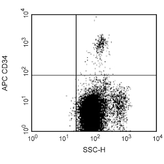
The 5E10 monoclonal antibody specifically binds to human CD90 which is also known as Thy-1. CD90 is a 25-35 kDa glycophosphatidylinositol-anchored membrane glycoprotein of the Ig superfamily that is expressed on 1-4% of human fetal liver cells, cord blood cells, and bone marrow cells. The anti-CD90 antibody binds to a subset of immature CD34+ cells and a distinct subset of mature CD34- cells that are CD3+CD4+. The CD90+CD34+ population is highly enriched for cells capable of long-term culture. The anti-CD90 antibody is useful for enriching high proliferative potential colony-forming cells (HIPP-CFC) that are primative progenitor cells.

568496 Rev. 2
Format Details
Down Arrow Up Arrow
RY586
The BD Horizon RealYellow™ 586 (RY586) Dye is part of the BD family of yellow-green dyes. It is a small organic fluorochrome with an excitation maximum (Ex Max) at 565-nm and an emission maximum (Em Max) at 586-nm. Driven by BD innovation, RY586 can be used on both spectral and conventional cytometers and is designed to be excited by the Yellow-Green laser (561-nm) with minimal excitation by the 488-nm Blue laser. For conventional instruments equipped with a Yellow-Green laser (561-nm), RY586 can be used as an alternative to PE and we recommend using an optical filter centered near 586-nm (eg, a 586/15-nm bandpass filter). For spectral instruments equipped with a Yellow-Green laser (561-nm), it can be used in conjunction with PE. Compared to PE, RY586 is similar in brightness, minimal spillover into Blue detectors, and increased spillover into the 610/20-nm (PE-CF594) detector. Please ensure that your instrument configuration (lasers and optical filters) is appropriate for this dye.
altImg
RY586
Yellow-Green 561 nm
564 nm
586 nm
568496 Rev.2
Citations & References
Down Arrow Up Arrow

Development References (7)

  1. Baum CM, Weissman IL, Tsukamoto AS, Buckle AM, Peault B. Isolation of a candidate human hematopoietic stem-cell population. Proc Natl Acad Sci U S A. 1992; 89(7):2804-2808. (Biology). View Reference
  2. Bjornson-Hooper ZB, Fragiadakis GK, Spitzer MH, et al. A Comprehensive Atlas of Immunological Differences Between Humans, Mice, and Non-Human Primates.. Front Immunol. 2022; 13:867015. (Clone-specific: Flow cytometry). View Reference
  3. Craig W, Kay R, Cutler RL, Lansdorp PM. Expression of Thy-1 on human hematopoietic progenitor cells. J Exp Med. 1993; 177(5):1331-1342. (Immunogen: Flow cytometry, Immunoprecipitation, Western blot). View Reference
  4. Kishimoto T. Tadamitsu Kishimoto .. et al., ed. Leucocyte typing VI : white cell differentiation antigens : proceedings of the sixth international workshop and conference held in Kobe, Japan, 10-14 November 1996. New York: Garland Pub.; 1997.
  5. Knapp W. W. Knapp .. et al., ed. Leucocyte typing IV : white cell differentiation antigens. Oxford New York: Oxford University Press; 1989:1-1182.
  6. Lansdorp PM, Thomas TE. AP Gee, ed. Bone Marrow Processing and Purging. Boca Raton FL: CRC Press; 1991.
  7. Schlossman SF. Stuart F. Schlossman .. et al., ed. Leucocyte typing V : white cell differentiation antigens : proceedings of the fifth international workshop and conference held in Boston, USA, 3-7 November, 1993. Oxford: Oxford University Press; 1995.
View All (7) View Less
568496 Rev. 2

Please refer to Support Documents for Quality Certificates


Global - Refer to manufacturer's instructions for use and related User Manuals and Technical data sheets before using this products as described


Comparisons, where applicable, are made against older BD Technology, manual methods or are general performance claims.  Comparisons are not made against non-BD technologies, unless otherwise noted.

For Research Use Only. Not for use in diagnostic or therapeutic procedures.