Skip to main content Skip to navigation
RY586 Mouse Anti-Human CD73
Multiparameter flow cytometric analysis of CD73 expression on human peripheral blood leucocytes. Human whole blood was stained with either BD Horizon™ RY586 Mouse IgG1, κ Isotype Control (Cat. No. 568097; Left Plot) or BD Horizon™ RY586 Mouse Anti-Human CD73 antibody (Cat. No. 568439/568440; Right Plot). The erythrocytes were lysed with BD FACS™ Lysing Solution (Cat. No. 349202). The bivariate pseudocolor density plot showing the correlated expression of CD20 (or Ig Isotype control staining) versus side light-scatter (SSC) signals was derived from gated events with the forward and side light-scatter characteristics of intact leucocytes. Flow cytometry and data analysis were performed using a BD LSRFortessa™ X-20 Cell Analyzer System and FlowJo™ software.
RY586 Mouse Anti-Human CD73
Flow cytometric analysis of CD73 expression on human peripheral blood lymphocytes. Human whole blood was stained with either BD Horizon™ RY586 Mouse IgG1, κ Isotype Control (Cat. No. 568097; dashed line histogram) or BD Horizon™ RY586 Mouse Anti-Human CD73 antibody (Cat. No. 568439/568440; solid line histogram). Erythrocytes were lysed with BD FACS Lysing Solution (Cat. No. 349202). The fluorescence histogram showing CD73 expression (or Ig Isotype control staining) was derived from gated events with the forward and side light-scatter characteristics of intact lymphocytes. Flow cytometric analysis was performed using a BD LSRFortessa™ X-20 Cell Analyzer System and FlowJo™ software.
Multiparameter flow cytometric analysis of CD73 expression on human peripheral blood leucocytes. Human whole blood was stained with either BD Horizon™ RY586 Mouse IgG1, κ Isotype Control (Cat. No. 568097; Left Plot) or BD Horizon™ RY586 Mouse Anti-Human CD73 antibody (Cat. No. 568439/568440; Right Plot). The erythrocytes were lysed with BD FACS™ Lysing Solution (Cat. No. 349202). The bivariate pseudocolor density plot showing the correlated expression of CD20 (or Ig Isotype control staining) versus side light-scatter (SSC) signals was derived from gated events with the forward and side light-scatter characteristics of intact leucocytes. Flow cytometry and data analysis were performed using a BD LSRFortessa™ X-20 Cell Analyzer System and FlowJo™ software.
Flow cytometric analysis of CD73 expression on human peripheral blood lymphocytes. Human whole blood was stained with either BD Horizon™ RY586 Mouse IgG1, κ Isotype Control (Cat. No. 568097; dashed line histogram) or BD Horizon™ RY586 Mouse Anti-Human CD73 antibody (Cat. No. 568439/568440; solid line histogram). Erythrocytes were lysed with BD FACS Lysing Solution (Cat. No. 349202). The fluorescence histogram showing CD73 expression (or Ig Isotype control staining) was derived from gated events with the forward and side light-scatter characteristics of intact lymphocytes. Flow cytometric analysis was performed using a BD LSRFortessa™ X-20 Cell Analyzer System and FlowJo™ software.
Product Details
Down Arrow Up Arrow


BD Horizon™
NT5E; 5' nucleotidase; 5'-NT; E5NT; Ecto-5'-nucleotidase; eN; eNT; NT; NT5
Human (QC Testing)
Mouse IgG1, κ
Pre-B leukemia cell line
Flow cytometry (Routinely Tested)
5 µl
V B-CD73.3
4907
Aqueous buffered solution containing ≤0.09% sodium azide.
RUO


Preparation And Storage

Store undiluted at 4°C and protected from prolonged exposure to light. Do not freeze. The monoclonal antibody was purified from tissue culture supernatant or ascites by affinity chromatography. The antibody was conjugated to the dye under optimum conditions and unreacted dye was removed.

Recommended Assay Procedures

BD® CompBeads can be used as surrogates to assess fluorescence spillover (compensation). When fluorochrome conjugated antibodies are bound to BD® CompBeads, they have spectral properties very similar to cells. However, for some fluorochromes there can be small differences in spectral emissions compared to cells, resulting in spillover values that differ when compared to biological controls. It is strongly recommended that when using a reagent for the first time, users compare the spillover on cells and BD® CompBeads to ensure that BD® CompBeads are appropriate for your specific cellular application.

Product Notices

  1. Please refer to www.bdbiosciences.com/us/s/resources for technical protocols.
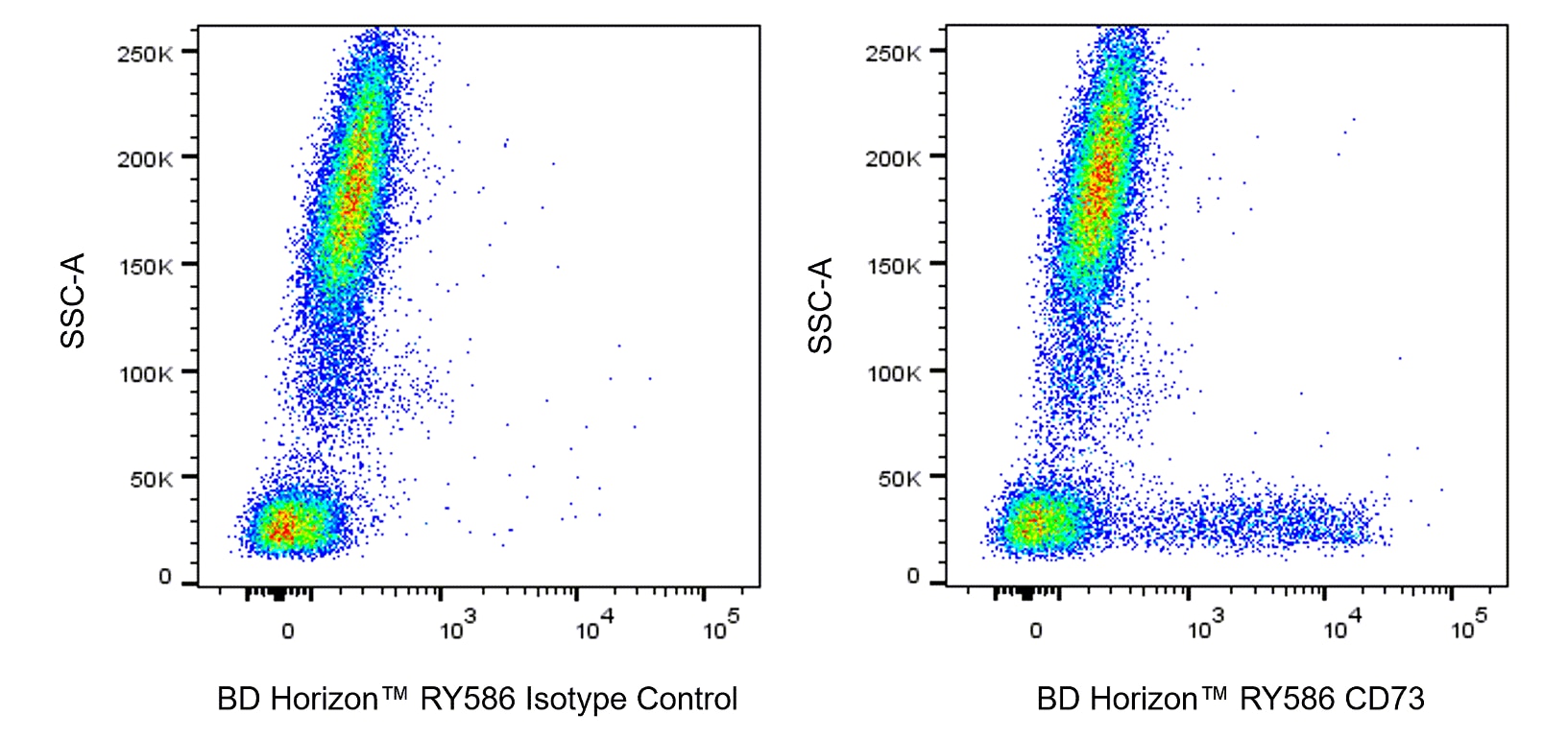
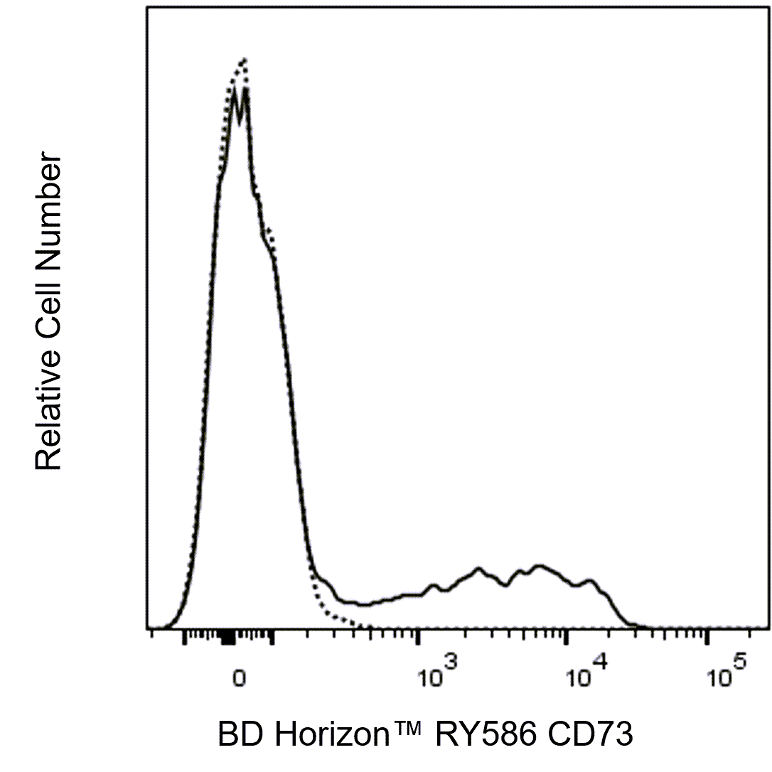
  2. This reagent has been pre-diluted for use at the recommended Volume per Test. We typically use 1 × 10^6 cells in a 100-µl experimental sample (a test).
  3. An isotype control should be used at the same concentration as the antibody of interest.
  4. Caution: Sodium azide yields highly toxic hydrazoic acid under acidic conditions. Dilute azide compounds in running water before discarding to avoid accumulation of potentially explosive deposits in plumbing.
  5. For fluorochrome spectra and suitable instrument settings, please refer to our Multicolor Flow Cytometry web page at www.bdbiosciences.com/colors.
  6. Human donor specific background has been observed in relation to the presence of anti-polyethylene glycol (PEG) antibodies, developed as a result of certain vaccines containing PEG, including some COVID-19 vaccines. We recommend use of BD Horizon Brilliant™ Stain Buffer in your experiments to help mitigate potential background. For more information visit https://www.bdbiosciences.com/en-us/support/product-notices.
  7. Please refer to http://regdocs.bd.com to access safety data sheets (SDS).
  8. CF™ is a trademark of Biotium, Inc.
568439 Rev. 1
Antibody Details
Down Arrow Up Arrow
AD2

The AD2 monoclonal antibody specifically binds to ecto-5'-nucleotidase, a 70 kDa, glycosyl phosphatidylinositol (GPI)-anchored glycoprotein. CD73 is expressed on subsets of T and B lymphocytes, follicular dendritic cells, epithelial cells, endothelial cells and mesenchymal stem cells. Its expression on lymphocytes increases during T and B cell development. CD73 has enzymatic activity and catalyzes the dephosphorylation of adenosine monophosphate (AMP) converting it to adenosine. It has been suggested that CD73 can mediate costimulatory signals in T cell activation and adhesion of lymphocytes to endothelium.

568439 Rev. 1
Format Details
Down Arrow Up Arrow
RY586
The BD Horizon RealYellow™ 586 (RY586) Dye is part of the BD family of yellow-green dyes. It is a small organic fluorochrome with an excitation maximum (Ex Max) at 565-nm and an emission maximum (Em Max) at 586-nm. Driven by BD innovation, RY586 can be used on both spectral and conventional cytometers and is designed to be excited by the Yellow-Green laser (561-nm) with minimal excitation by the 488-nm Blue laser. For conventional instruments equipped with a Yellow-Green laser (561-nm), RY586 can be used as an alternative to PE and we recommend using an optical filter centered near 586-nm (eg, a 586/15-nm bandpass filter). For spectral instruments equipped with a Yellow-Green laser (561-nm), it can be used in conjunction with PE. Compared to PE, RY586 is similar in brightness, minimal spillover into Blue detectors, and increased spillover into the 610/20-nm (PE-CF594) detector. Please ensure that your instrument configuration (lasers and optical filters) is appropriate for this dye.
altImg
RY586
Yellow-Green 561 nm
564 nm
586 nm
568439 Rev.1
Citations & References
Down Arrow Up Arrow

Development References (6)

  1. Alam MS, Kurtz CC, Rowlett RM, et al. CD73 is expressed by human regulatory T helper cells and suppresses proinflammatory cytokine production and Helicobacter felis-induced gastritis in mice. J Infect Dis. 2009; 199(4):494-504. (Biology). View Reference
  2. Dörken B, Möller P, Pezzutto R, Schwartz-Albiez R, Moldenhauer G. B-cell antigens: CD73. In: Knapp W. W. Knapp .. et al., ed. Leucocyte typing IV : white cell differentiation antigens. Oxford New York: Oxford University Press; 1989:102-104.
  3. Nakamura T, Kubagawa H, Ohno T, Cooper MD. Characterization of an IgM Fc-binding receptor on human T cells. J Immunol. 1993; 151(12):6933-6941. (Clone-specific). View Reference
  4. Salazar-Gonzalez JF, Moody DJ, Giorgi JV, Martinez-Maza O, Mitsuyasu RT, Fahey JL. Reduced ecto-5'-nucleotidase activity and enhanced OKT10 and HLA-DR expression on CD8 (T suppressor/cytotoxic) lymphocytes in the acquired immune deficiency syndrome: evidence of CD8 cell immaturity. J Immunol. 1985; 135(3):1778-1785. (Biology). View Reference
  5. Schlossman SF. Stuart F. Schlossman .. et al., ed. Leucocyte typing V : white cell differentiation antigens : proceedings of the fifth international workshop and conference held in Boston, USA, 3-7 November, 1993. Oxford: Oxford University Press; 1995.
  6. Thomson LF, Ruedi JM, Glass A, et al. Production and characterization of monoclonal antibodies to the glycosyl phosphatidylinositol-anchored lymphocyte differentiation antigen ecto-5'-nucleotidase (CD73). Tissue Antigens. 1990; 35(1):9-19. (Biology). View Reference
View All (6) View Less
568439 Rev. 1

Please refer to Support Documents for Quality Certificates


Global - Refer to manufacturer's instructions for use and related User Manuals and Technical data sheets before using this products as described


Comparisons, where applicable, are made against older BD Technology, manual methods or are general performance claims.  Comparisons are not made against non-BD technologies, unless otherwise noted.

For Research Use Only. Not for use in diagnostic or therapeutic procedures.