Skip to main content Skip to navigation
RY586 Mouse Anti-Human CD45RA
Two-color flow cytometric analysis of CD45RA expression on Human peripheral blood lymphocytes. Human whole blood was stained with BD Horizon™ BV421 Mouse Anti-Human CD45RO antibody (Cat. No. 562641/562649) and with either BD Horizon™ RY586 Mouse IgG2b, κ Isotype Control (Cat. No. 568132​; Left Plot) or BD Horizon™ RY586 Mouse Anti-Human CD45RA antibody (Cat. No. 568445/568446; Right Plot). The erythrocytes were lysed with BD FACS™ Lysing Solution (Cat. No. 349202). The bivariate pseudocolor density plot showing the correlated expression of CD45RA versus CD45RO was derived from gated events with the forward and side light-scatter characteristics of intact lymphocytes. Flow cytometry and data analysis were performed using a BD LSRFortessa™ X-20 Cell Analyzer System and FlowJo™ software.
Product Details
Down Arrow Up Arrow


BD Horizon™
CD45R; PTPRC; LCA; Leukocyte common antigen
Human (QC Testing)
Mouse IgG2b, κ
Flow cytometry (Routinely Tested)
5 µl
IV N906
5788
Aqueous buffered solution containing ≤0.09% sodium azide.
RUO


Preparation And Storage

Store undiluted at 4°C and protected from prolonged exposure to light. Do not freeze. The monoclonal antibody was purified from tissue culture supernatant or ascites by affinity chromatography. The antibody was conjugated to the dye under optimum conditions and unreacted dye was removed.

Recommended Assay Procedures

BD® CompBeads can be used as surrogates to assess fluorescence spillover (compensation). When fluorochrome conjugated antibodies are bound to BD® CompBeads, they have spectral properties very similar to cells. However, for some fluorochromes there can be small differences in spectral emissions compared to cells, resulting in spillover values that differ when compared to biological controls. It is strongly recommended that when using a reagent for the first time, users compare the spillover on cells and BD® CompBeads to ensure that BD® CompBeads are appropriate for your specific cellular application.

Product Notices

  1. Please refer to www.bdbiosciences.com/us/s/resources for technical protocols.
  2. This reagent has been pre-diluted for use at the recommended Volume per Test. We typically use 1 × 10^6 cells in a 100-µl experimental sample (a test).
  3. An isotype control should be used at the same concentration as the antibody of interest.
  4. Caution: Sodium azide yields highly toxic hydrazoic acid under acidic conditions. Dilute azide compounds in running water before discarding to avoid accumulation of potentially explosive deposits in plumbing.
  5. For fluorochrome spectra and suitable instrument settings, please refer to our Multicolor Flow Cytometry web page at www.bdbiosciences.com/colors.
  6. Human donor specific background has been observed in relation to the presence of anti-polyethylene glycol (PEG) antibodies, developed as a result of certain vaccines containing PEG, including some COVID-19 vaccines. We recommend use of BD Horizon Brilliant™ Stain Buffer in your experiments to help mitigate potential background. For more information visit https://www.bdbiosciences.com/en-us/support/product-notices.
  7. CF™ is a trademark of Biotium, Inc.
  8. Please refer to http://regdocs.bd.com to access safety data sheets (SDS).
568445 Rev. 1
Antibody Details
Down Arrow Up Arrow
HI100

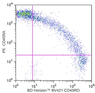
The HI100 monoclonal antibody specifically binds to the 220 kDa isoform of the human leukocyte common antigen, CD45RA. CD45RA is expressed on approximately 40-50% of peripheral CD4+ T cells, 50% of peripheral CD8+ T cells and on a portion of B cells and monocytes. The CD45RA antigen is expressed by naïve and activated T cells. CD45RA-specific antibodies are useful for the study of the suppressor/inducer subpopulation of CD4+ lymphocytes.

568445 Rev. 1
Format Details
Down Arrow Up Arrow
RY586
The BD Horizon RealYellow™ 586 (RY586) Dye is part of the BD family of yellow-green dyes. It is a small organic fluorochrome with an excitation maximum (Ex Max) at 565-nm and an emission maximum (Em Max) at 586-nm. Driven by BD innovation, RY586 can be used on both spectral and conventional cytometers and is designed to be excited by the Yellow-Green laser (561-nm) with minimal excitation by the 488-nm Blue laser. For conventional instruments equipped with a Yellow-Green laser (561-nm), RY586 can be used as an alternative to PE and we recommend using an optical filter centered near 586-nm (eg, a 586/15-nm bandpass filter). For spectral instruments equipped with a Yellow-Green laser (561-nm), it can be used in conjunction with PE. Compared to PE, RY586 is similar in brightness, minimal spillover into Blue detectors, and increased spillover into the 610/20-nm (PE-CF594) detector. Please ensure that your instrument configuration (lasers and optical filters) is appropriate for this dye.
altImg
RY586
Yellow-Green 561 nm
564 nm
586 nm
568445 Rev.1
Citations & References
Down Arrow Up Arrow

Development References (3)

  1. Knapp W. W. Knapp .. et al., ed. Leucocyte typing IV : white cell differentiation antigens. Oxford New York: Oxford University Press; 1989:1-1182.
  2. Koristka S, Cartellieri M, Theil A, et al. Retargeting of human regulatory T cells by single-chain bispecific antibodies.. J Immunol. 2012; 188(3):1551-8. (Clone-specific: Flow cytometry, Fluorescence activated cell sorting). View Reference
  3. Zola H. Leukocyte and stromal cell molecules : the CD markers. Hoboken, N.J.: Wiley-Liss; 2007.
568445 Rev. 1

Please refer to Support Documents for Quality Certificates


Global - Refer to manufacturer's instructions for use and related User Manuals and Technical data sheets before using this products as described


Comparisons, where applicable, are made against older BD Technology, manual methods or are general performance claims.  Comparisons are not made against non-BD technologies, unless otherwise noted.

For Research Use Only. Not for use in diagnostic or therapeutic procedures.