Preparation And Storage
Recommended Assay Procedures
BD® CompBeads can be used as surrogates to assess fluorescence spillover (compensation). When fluorochrome conjugated antibodies are bound to BD® CompBeads, they have spectral properties very similar to cells. However, for some fluorochromes there can be small differences in spectral emissions compared to cells, resulting in spillover values that differ when compared to biological controls. It is strongly recommended that when using a reagent for the first time, users compare the spillover on cells and BD® CompBeads to ensure that BD® CompBeads are appropriate for your specific cellular application.
Product Notices
- Please refer to www.bdbiosciences.com/us/s/resources for technical protocols.
- Caution: Sodium azide yields highly toxic hydrazoic acid under acidic conditions. Dilute azide compounds in running water before discarding to avoid accumulation of potentially explosive deposits in plumbing.
- This reagent has been pre-diluted for use at the recommended Volume per Test. We typically use 1 × 10^6 cells in a 100-µl experimental sample (a test).
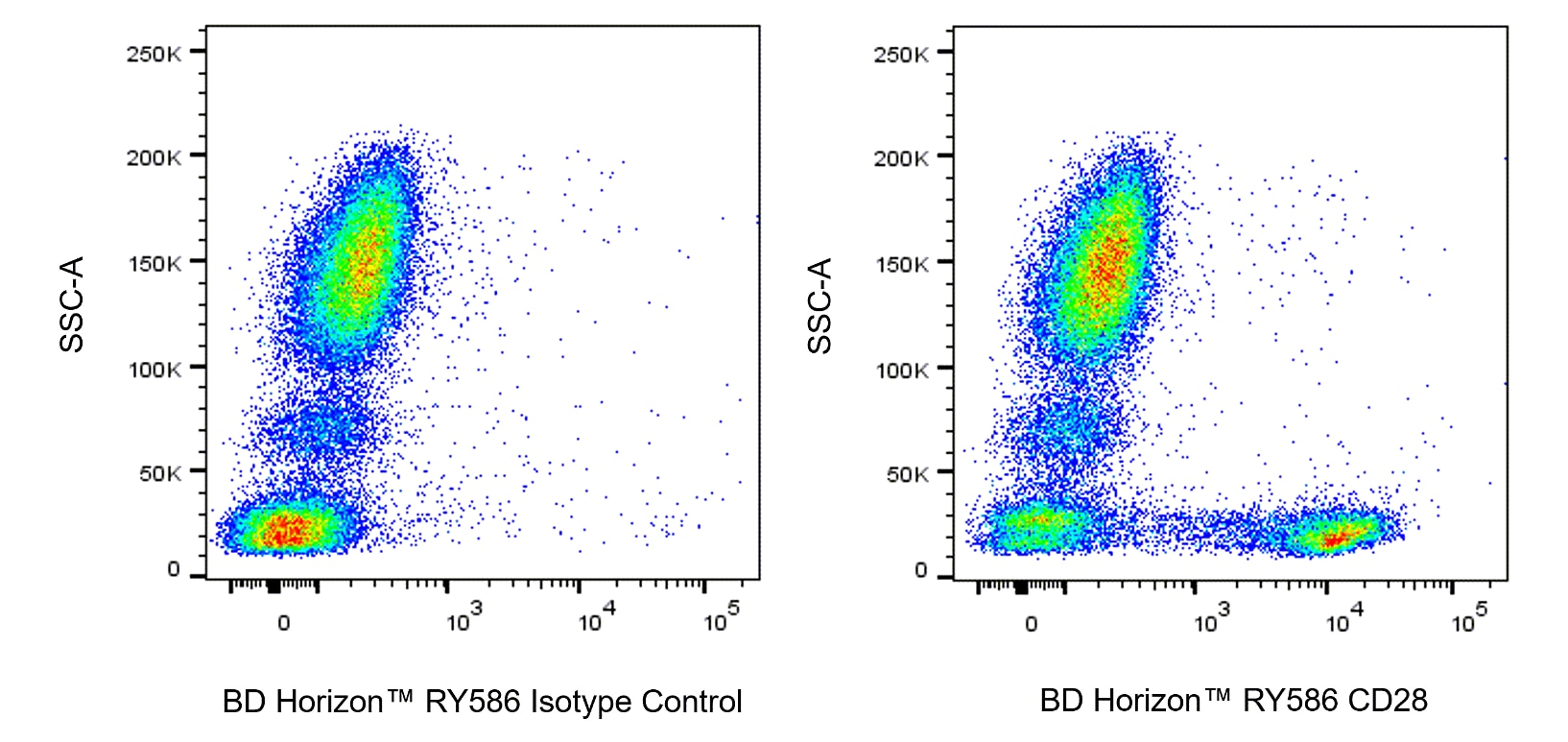
- For fluorochrome spectra and suitable instrument settings, please refer to our Multicolor Flow Cytometry web page at www.bdbiosciences.com/colors.
- An isotype control should be used at the same concentration as the antibody of interest.
- Species cross-reactivity detected in product development may not have been confirmed on every format and/or application.
- Human donor specific background has been observed in relation to the presence of anti-polyethylene glycol (PEG) antibodies, developed as a result of certain vaccines containing PEG, including some COVID-19 vaccines. We recommend use of BD Horizon Brilliant™ Stain Buffer in your experiments to help mitigate potential background. For more information visit https://www.bdbiosciences.com/en-us/support/product-notices.
- Please refer to http://regdocs.bd.com to access safety data sheets (SDS).
- CF™ is a trademark of Biotium, Inc.
Companion Products


The CD28.2 monoclonal antibody specifically binds to CD28, a 44 kDa homodimeric transmembrane glycoprotein present on most mature T cells, thymocytes and plasma cells. CD28 is a costimulatory receptor that binds CD80 and CD86 as ligands and plays a very important role in T cell-B cell interactions. It has been suggested that CD28 initiates and regulates a separate and distinct signal transduction pathway from those stimulated by the TCR complex. Additionally, it has been reported that CD28 antibody clones vary in their ability to stimulate T cells to produce IL-2 and increase intracellular Ca2+ concentration. This finding suggests the existence of functionally distinct subregions on the CD28 molecule. CD28.2 has been demonstrated to bind to the same molecule as clone L293, another CD28 mAb, and has been reported to induce Ca2+ influx in Jurkat T cells.

Development References (7)
-
Barclay NA, Brown MH, Birkeland ML, et al, ed. The Leukocyte Antigen FactsBook. San Diego, CA: Academic Press; 1997.
-
June CH, Bluestone JA, Nadler LM, Thompson CB. The B7 and CD28 receptor families. Immunol Today. 1994; 15(7):321-331. (Biology). View Reference
-
Kuiper H, Brouwer M, Vermeire S, van Lier R. Analysis of the Workshop CD28 Panel mAb: distinct signalling pathways coupled to CD28. In: Schlossman SF. Stuart F. Schlossman .. et al., ed. Leucocyte typing V : white cell differentiation antigens : proceedings of the fifth international workshop and conference held in Boston, USA, 3-7 November, 1993. Oxford: Oxford University Press; 1995:373-374.
-
Nunes J, Klasen S, Franco MD, et al. Signalling through CD28 T-cell activation pathway involves an inositol phospholipid-specific phospholipase C activity. Biochem J. 1993; 293(3):835-842. (Clone-specific: Calcium Flux, (Co)-stimulation, Functional assay). View Reference
-
Nunes J, Klasen S, Ragueneau M, et al. CD28 mAbs with distinct binding properties differ in their ability to induce T cell activation: analysis of early and late activation events. Int Immunol. 1993; 5(3):311-315. (Immunogen: Calcium Flux, (Co)-stimulation, Flow cytometry, Functional assay, IC/FCM Block, Immunoprecipitation, Stimulation). View Reference
-
Olive D, Cerdan C, Costello R, et al. CD28 and CTLA-4 cluster report. In: Schlossman SF. Stuart F. Schlossman .. et al., ed. Leucocyte typing V : white cell differentiation antigens : proceedings of the fifth international workshop and conference held in Boston, USA, 3-7 November, 1993. Oxford: Oxford University Press; 1995:360-370.
-
Thaker YR, Schneider H, Rudd CE. TCR and CD28 activate the transcription factor NF-κB in T-cells via distinct adaptor signaling complexes.. Immunol Lett. 2015; 163(1):113-9. (Clone-specific: Activation, Flow cytometry). View Reference
Please refer to Support Documents for Quality Certificates
Global - Refer to manufacturer's instructions for use and related User Manuals and Technical data sheets before using this products as described
Comparisons, where applicable, are made against older BD Technology, manual methods or are general performance claims. Comparisons are not made against non-BD technologies, unless otherwise noted.
For Research Use Only. Not for use in diagnostic or therapeutic procedures.